. 2017 Mar 24:7:44847.
doi: 10.1038/srep44847.

The miR-24-3p/p130Cas: a novel axis regulating the migration and invasion of cancer cells

Affiliations

The miR-24-3p/p130Cas: a novel axis regulating the migration and invasion of cancer cells

Hoin Kang et al. Sci Rep. .

Erratum in

Abstract

MicroRNAs (miRNAs) are small non-coding RNAs that negatively regulate gene expression by suppressing translation or facilitating mRNA decay. Differential expression of miRNAs is involved in the pathogenesis of several diseases including cancer. Here, we investigated the role of-miR-24-3p as a downregulated miRNA in metastatic cancer. miR-24-3p was decreased in metastatic cancer and lower expression of miR-24-3p was related to poor survival of cancer patients. Consistently, ectopic expression of miR-24-3p suppressed the cell migration, invasion, and proliferation of MCF7, Hep3B, B16F10, SK-Hep1, and PC-3 cells by directly targeting p130Cas. Stable expression of p130Cas restored miR-24-3p-mediated inhibition of cell migration and invasion. These results suggest that miR-24-3p functions as a tumor suppressor and the miR-24-3p/p130Cas axis is a novel factor of cancer progression by regulating cell migration and invasion.


Conflict of interest statement

The authors declare no competing financial interests.

Figures
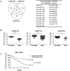
Figure 1
Figure 1. Differential expression of miR-24-3p in metastatic cancer.
(A) Analysis of differentially expressed miRNAs in metastatic cancer using three different Gene Expression Omnibus databases (GES67138 and GES67139, human hepatocellular carcinoma; GES21306, human prostate cancer). The number indicates the downregulated miRNAs in metastatic cancers and the list of miRNAs is shown as a table. (B) The relative level of miR-24-3p in the metastatic group and the non-metastatic group was analyzed from three different sets of microRNA microarray data. (C) Kaplan-Meier survival curve. The relation between the overall survival rate of cancer patients and the miR-24-3p level was analyzed in the GES31384 dataset.
Figure 2
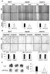
Figure 2. miR-24-3p suppressed migration and invasion of cancer cells.
(A) Analysis of wound closure after miRNA transfection. MCF7, Hep3B, and B16F10 cells were transfected with either miR-24-3p mimic or control miRNAs and cultured until they reached confluency. After wounds were created, the cell migration distance was analyzed 48 h later. (B) Migration and invasion assay. After the transfection of miRNAs, cells were cultured in transwell with or without matrigel, and migrated cells were stained and analyzed by counting cells from three different fields. (C) After transfection of miR-24-3p and control miRNA, B16F10 cells (3 × 105 cells/mouse) were injected into the tail vein of C57BL6 mice (n = 5). 17 days later, mice were sacrificed and the lungs were isolated. The number of B16F10 colonies present on the surface of each set of lungs was determined by visual inspection. Data are representative from three independent experiments. *p < 0.05.
Figure 3
Figure 3. miR-24-3p suppressed growth of cancer cells.
(A) After the transfection of miRNAs, MCF7, Hep3B, and B16F10 cells were cultured with or without the anti-cancer drugs including tamoxifen, 5-FU, CDDP, and doxorubicin, and cell viability was assessed by MTT assay. (B) MCF7 cells transfected with either miR-24-3p or control miRNA and 1 × 103 cells were cultured for 3 weeks to form colonies. Colonies were fixed and stained with cystal violet solution and the number of colonies was counted. (C) miRNA-transfected MCF7 cells were transplanted into the mice (n = 5) and tumor development was observed over 4 weeks. Xenograft tumor mass and relative miRNA levels in tumors were analyzed from 4 different mice. Data are representative or indicate the mean ± SEM of three independent experiments. *p < 0.05; **p < 0.01.
Figure 4
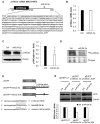
Figure 4. miR-24-3p down-regulated p130Cas expression.
(A) Schematic diagram of p130Cas mRNA with miR-24-3p binding site. The predicted miR-24-3p binding site was found in the 3′UTR of p130Cas mRNA (underlined). (B and C) After the transfection of miRNAs, p130Cas mRNA and protein levels were determined by RT-qPCR (B) and Western blotting (C), respectively. GAPDH mRNA or β-actin was used as the internal control. (D) Translational regulation of p130Cas by miR-24-3p. After the incubation of miRNA-transfected MCF7 cells with 35S-methionine/cysteine, 35S-p130Cas protein was isolated using p130Cas antibody, separated by SDS-PAGE and visualized using PharoseFX Plus. The representative image shows relative levels of nascent p130Cas. (E) A schematic of the EGFP-reporter constructs. The 3′UTR of p130Cas mRNA containing a miR-24-3p binding site (3002–3150 nt) was inserted into pEGFP-C1 and a mutant reporter construct lacking the miR-binding site was generated using site-directed mutagenesis. (F) Reporter analysis after the miR-24-3p expression. After transfection of MCF7 cells with miRs and reporter constructs, EGFP levels were evaluated by Western blotting. β-actin was used as a loading control. All data are presented as mean ± SD of three independent experiments. *p < 0.05.
Figure 5
Figure 5. miR-24-3p suppressed cancer progression via p130Cas regulation.
(A and B) MCF7_Control and MCF7_EGFP-Cas cells were transfected with either miR-24-3p mimic or control miRNAs and cell migration/invasion was analyzed. After the cells reached confluency, wound closure was determined by measuring cell migration distance (A). Cells were cultured in transwell with or without matrigel, and migrated cells were stained and analyzed by counting cells from three different fields (B). (C) 1 × 103 of miRNA-transfected cells were cultured for 3 weeks. The colonies were stained with crystal violet and counted in 3 randomly selected visual fields. The images were representative of three independent experiments and graphs indicate the mean ± SEM of three independent experiments. *p < 0.05.

Similar articles

Cited by

References

    1. Collins C. & Nelson W. J. Running with neighbors: coordinating cell migration and cell-cell adhesion. Current opinion in cell biology 36, 62–70, doi: 10.1016/j.ceb.2015.07.004 (2015). - DOI - PMC - PubMed
    1. Vieira A. F. & Paredes J. P-cadherin and the journey to cancer metastasis. Molecular cancer 14, 178, doi: 10.1186/s12943-015-0448-4 (2015). - DOI - PMC - PubMed
    1. Eke I. & Cordes N. Focal adhesion signaling and therapy resistance in cancer. Seminars in cancer biology 31, 65–75, doi: 10.1016/j.semcancer.2014.07.009 (2015). - DOI - PubMed
    1. Won Y. K. et al.. Stereotactic radiosurgery for brain metastasis in non-small cell lung cancer. Radiation oncology journal 33, 207–216, doi: 10.3857/roj.2015.33.3.207 (2015). - DOI - PMC - PubMed
    1. Bartel D. P. MicroRNAs: genomics, biogenesis, mechanism, and function. Cell 116, 281–297 (2004). - PubMed

Publication types

LinkOut - more resources