. 2017 Mar 24:7:45152.
doi: 10.1038/srep45152.

Intermediate filament reorganization dynamically influences cancer cell alignment and migration

Affiliations

Intermediate filament reorganization dynamically influences cancer cell alignment and migration

Andrew W Holle et al. Sci Rep. .

Abstract

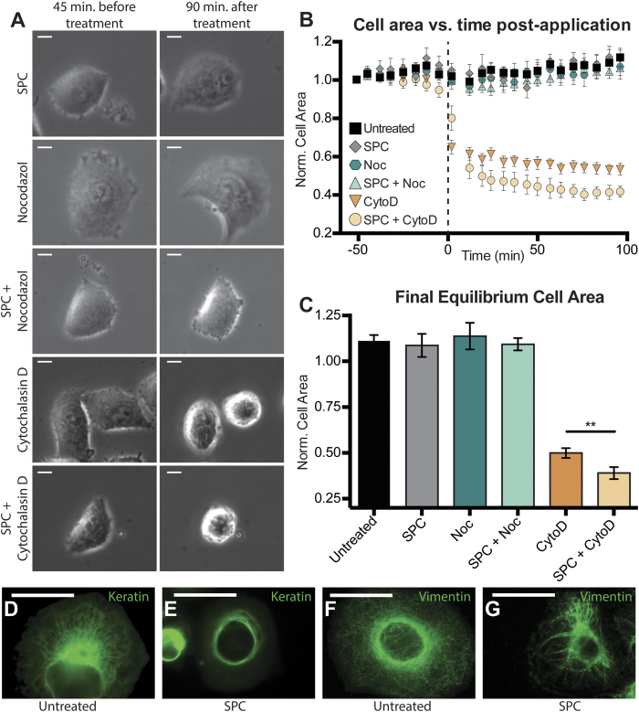
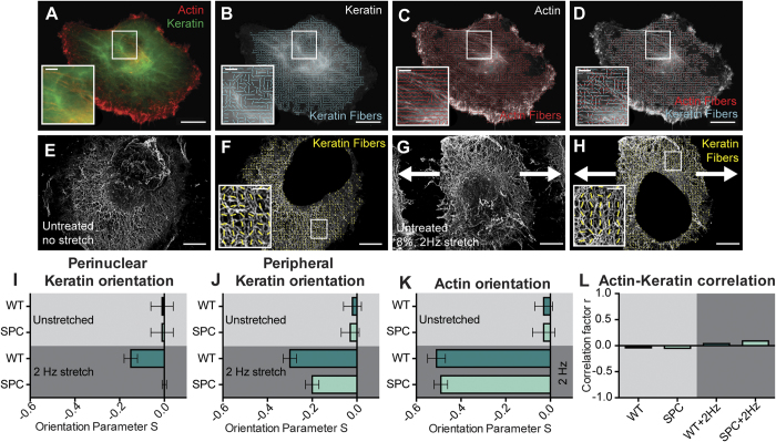
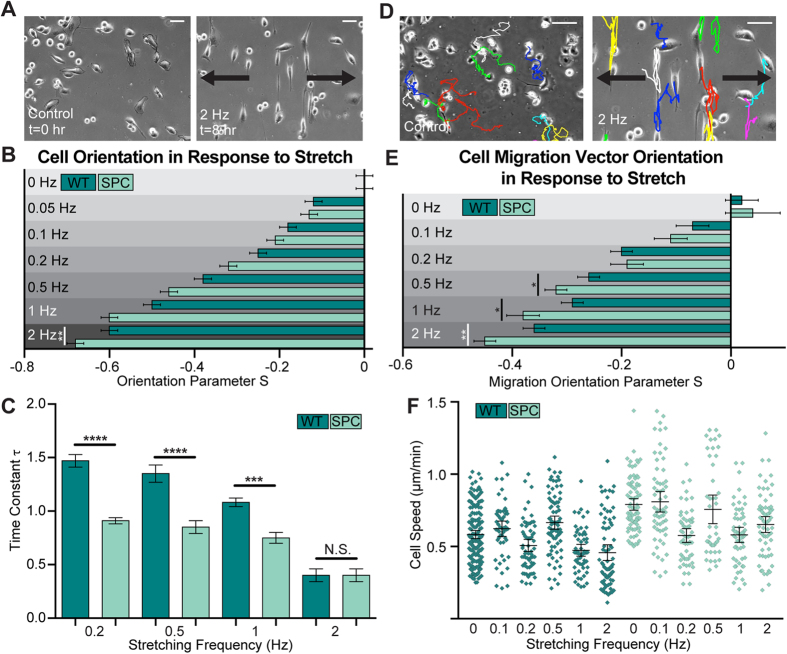
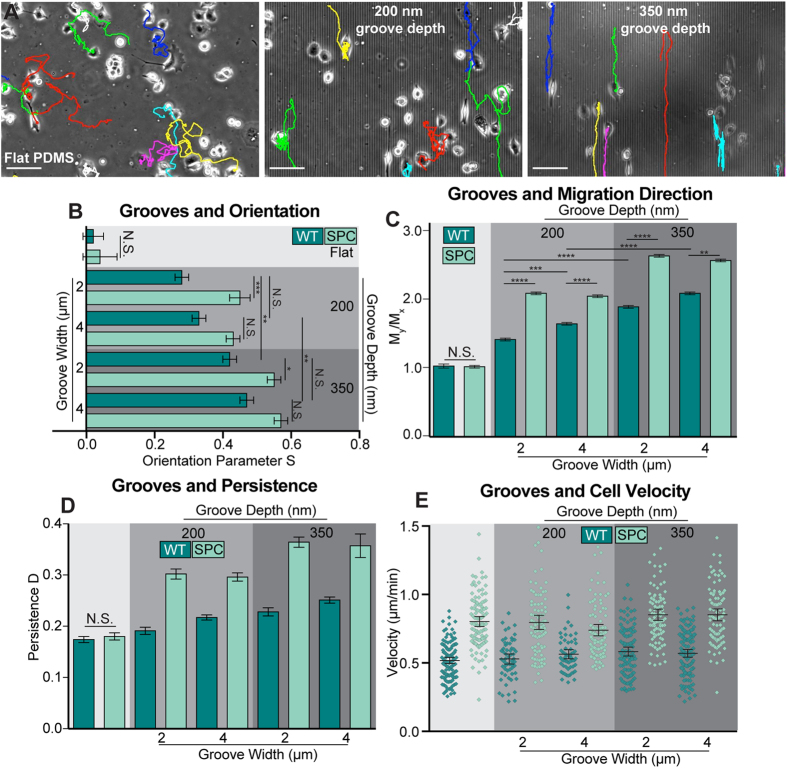
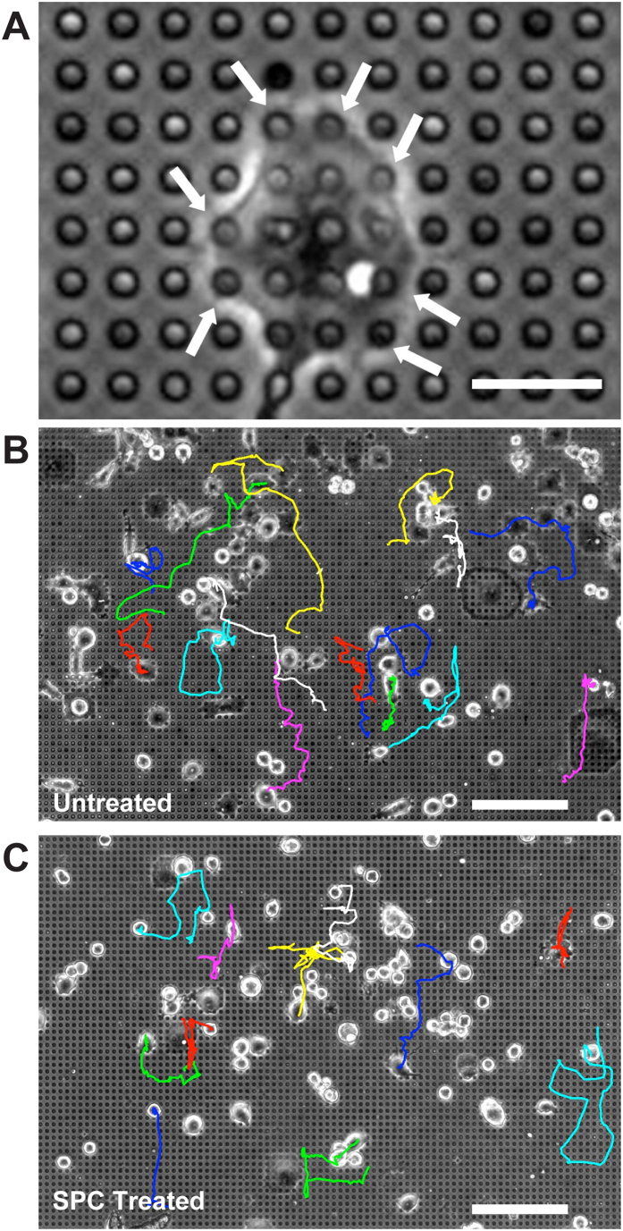
The interactions between a cancer cell and its extracellular matrix (ECM) have been the focus of an increasing amount of investigation. The role of the intermediate filament keratin in cancer has also been coming into focus of late, but more research is needed to understand how this piece fits in the puzzle of cytoskeleton-mediated invasion and metastasis. In Panc-1 invasive pancreatic cancer cells, keratin phosphorylation in conjunction with actin inhibition was found to be sufficient to reduce cell area below either treatment alone. We then analyzed intersecting keratin and actin fibers in the cytoskeleton of cyclically stretched cells and found no directional correlation. The role of keratin organization in Panc-1 cellular morphological adaptation and directed migration was then analyzed by culturing cells on cyclically stretched polydimethylsiloxane (PDMS) substrates, nanoscale grates, and rigid pillars. In general, the reorganization of the keratin cytoskeleton allows the cell to become more 'mobile'- exhibiting faster and more directed migration and orientation in response to external stimuli. By combining keratin network perturbation with a variety of physical ECM signals, we demonstrate the interconnected nature of the architecture inside the cell and the scaffolding outside of it, and highlight the key elements facilitating cancer cell-ECM interactions.


Conflict of interest statement

The authors declare no competing financial interests.

Figures

Figure 1
Figure 1. Keratin Phosphorylation and Cytoskeletal Inhibitors.
(A) Images of single cells were taken both 45 minutes before and 90 minutes after application of inhibitors or SPC. (B) Average normalized cell area was calculated from the time of application to equilibrium (~90 minutes) and (C) final equilibrium values for normalized cell area for each treatment showed a significant difference between Cytochalasin D treatment alone and treatment in conjunction with keratin phosphorylation (**p < 0.01 by ANOVA). Untreated keratin and vimentin distribution (D,F) was altered upon application of SPC and resulted in perinuclear organization (E,G). Scale bars = 50 μm (A,D,E). SPC- Sphingosylphosphorylcholine, Noc- Nocodazole, CytoD- Cytochalasin D.
Figure 2
Figure 2. Actin and Keratin Alignment in Response to Cyclic Strain.
(A) Immunofluorescence image example used to calculate keratin (B) and actin (C) alignment, followed by alignment correlation analysis of intersecting fibers (D). Electron microscopy was used to visualize peripheral keratin alignment on both unstrained and strained substrates (EH). On unstretched substrates, keratin and actin did not display any non-random orientation (IK, top), but upon application of 2 Hz cyclic substrate strain, all fibers realigned in a perpendicular direction, with actin responding most strongly (IK, bottom). Despite both fibers realigning, local alignment of actin and keratin did not correlate strongly in the cyclically strained samples (L). Scale bars = 20 μm (AD), 5 μm (AD insets, EF), 1 μm (F,H insets). WT = Untreated (IL). Arrows indicate direction of cyclic strain (G,H).
Figure 3
Figure 3. Keratin Phosphorylation and Cyclic Strain Induced Orientation and Migration.
(A) Images of single cells were taken over the course of 8 hours during the application of various cyclic strain frequencies, with most cells aligning most efficiently perpendicular to the direction of strain at higher frequencies and with SPC treatment (B). Analysis of the amount of time needed to achieve orientation equilibrium revealed that SPC can augment rearrangement speed in frequencies up to 2 Hz, where the effect is lost (C). By tracking cells during the experiment (D), migration direction vectors could be calculated and analyzed in the context of alignment (E), with higher strain frequencies and SPC treatment both contributing to more directed migration. SPC treatment caused the cells to move faster, but strain frequency had no systemic effect on cell migration speed (F). Increases in cell speed were statistically significant for control substrates (p < 0.0001), 0.2 Hz (p < 0.0001), 0.5 Hz (p < 0.01), and 2 Hz substrates (p < 0.0001), all by one-way ANOVA with Tukey post hoc tests. Scale bars = 50 μm (A), 150 μm (D). WT = Untreated (BF). Arrows indicate direction of cyclic strain (A,D).
Figure 4
Figure 4. Keratin Phosphorylation and Groove Depth Induce Alignment and Directed Migration.
Images of cells on flat control substrates and grooved substrates with different dimensions were obtained, with deeper grooves causing stronger alignment and directed migration (A). Analysis of the cells on the grooved substrates revealed that groove width was not as important as groove depth for cell orientation (B), cell migration vector orientation (C), or migration persistence (D). While keratin phosphorylation caused an increase in cell velocity, substrate grooves did not alter the velocity profile of either the untreated or SPC treated cells (E). Cell speed increases as a result of SPC treatment were statistically significant (p < 0.0001) by one-way ANOVA with Tukey post hoc tests for all culture conditions. Scale bars = 150 μm (A). WT = Untreated (BE).
Figure 5
Figure 5. Keratin Phosphorylation and Influences Cell Migration on Pillar Substrates.
Panc-1 cells were plated onto substrates containing ECM protein atop pillars, confining adhesion to these points (A). Tracking cell migration over a period of eight hours revealed distinct migration patterns in (B) untreated and (C) SPC-treated cells. Scale bars = 30 μm (A), 150 μm (B,C).

References

    1. Wehrle-Haller B. & Imhof B. A. Actin, microtubules and focal adhesion dynamics during cell migration. The International Journal of Biochemistry & Cell Biology 35, 39–50 (2003). - PubMed
    1. Schoumacher M., Goldman R. D., Louvard D. & Vignjevic D. M. Actin, microtubules, and vimentin intermediate filaments cooperate for elongation of invadopodia. J. Cell Biol. 189, 541–556 (2010). - PMC - PubMed
    1. Gavara N., Roca-Cusachs P., Sunyer R., Farré R. & Navajas D. Mapping Cell-Matrix Stresses during Stretch Reveals Inelastic Reorganization of the Cytoskeleton. Biophys. J. 95, 464–471 (2008). - PMC - PubMed
    1. Pellegrin S. & Mellor H. Actin stress fibres. J Cell Sci 120, 3491–3499 (2007). - PubMed
    1. Pollard T. D. & Cooper J. A. Actin, a central player in cell shape and movement. Science 326, 1208–1212 (2009). - PMC - PubMed

Publication types

LinkOut - more resources