. 2017 Mar 24:7:45160.
doi: 10.1038/srep45160.

The Effect Of microbial Mats In The Decay Of Anurans With Implications For Understanding Taphonomic Processes In The Fossil Record

Affiliations

The Effect Of microbial Mats In The Decay Of Anurans With Implications For Understanding Taphonomic Processes In The Fossil Record

M Iniesto et al. Sci Rep. .

Abstract

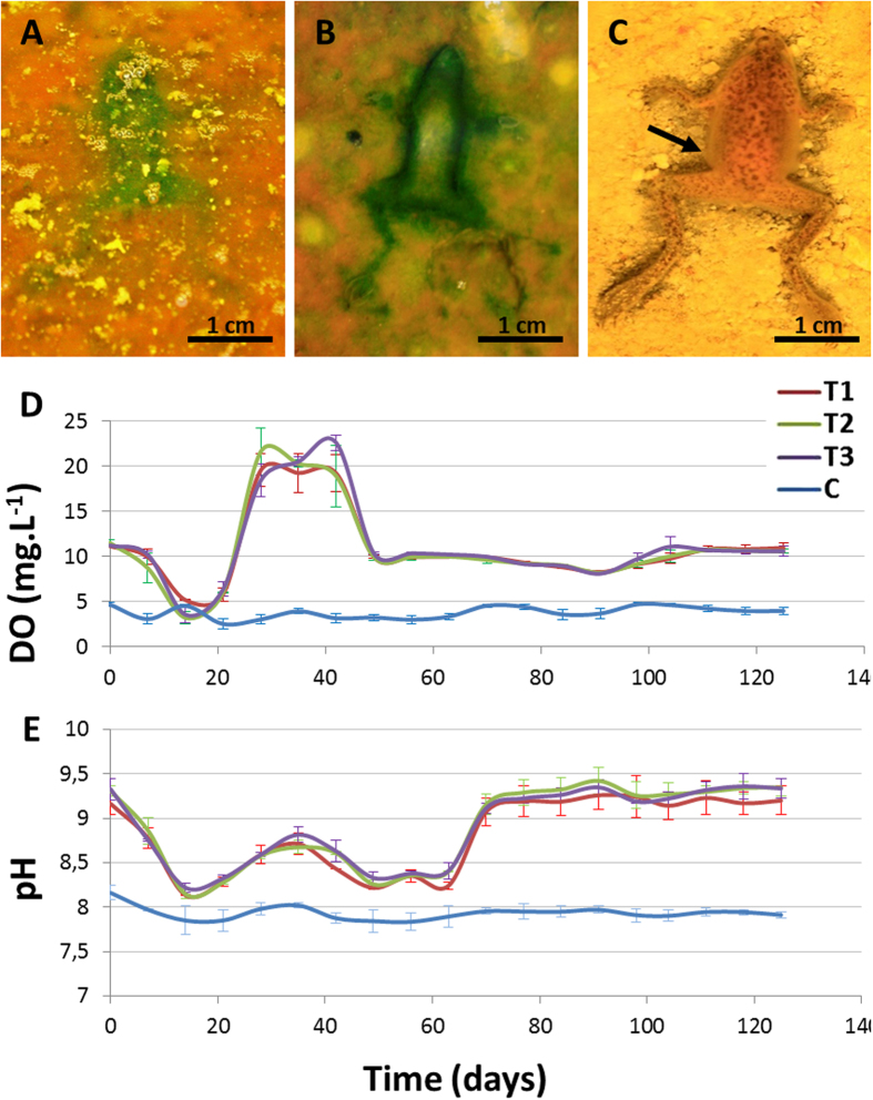
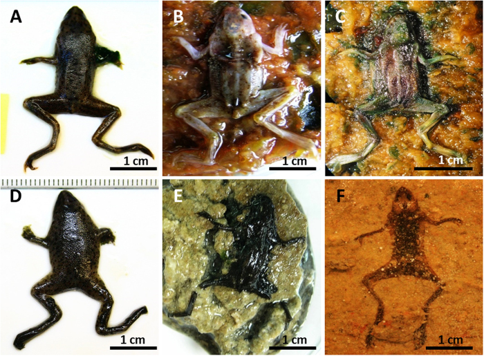
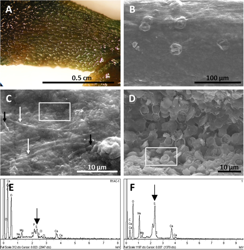
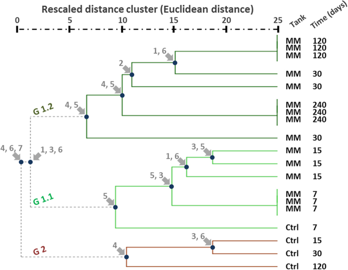
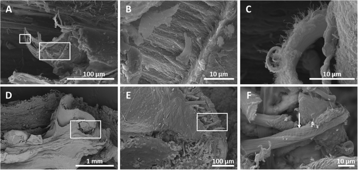
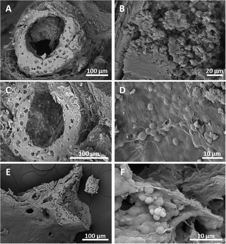
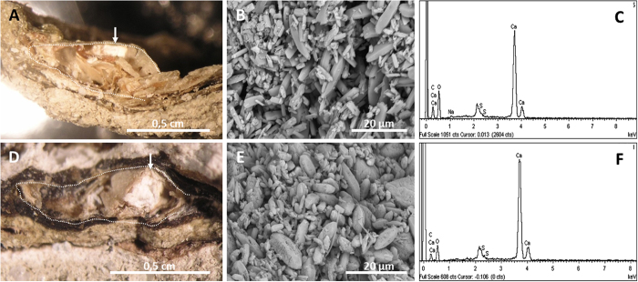
The pattern and sequence of the decomposition of the Pipidae African dwarf frog (Hymenochirus boettgeri) is tracked in an experiment with microbial mats in order to explore soft tissue preservation over three years. Frog decay in microbial mats is preceded by rapid entombment (25-30 days) and mediated by the formation of a sarcophagus, which is built by a complex microbial community. The frog carcasses maintained a variety of soft tissues for years. Labile organic structures show greater durability within the mat, cells maintain their general shape (bone marrow cells and adipocytes), and muscles and connective tissues (adipose and fibrous tendons) exhibit their original organic structures. In addition, other soft tissues are promptly mineralized (day 540) in a Ca-rich carbonate phase (encephalic tectum) or enriched in sulphur residues (integumentary system). The result is coherent with a bias in soft-tissue preservation, as some tissues are more likely to be conserved than others. The outcomes support observations of exceptionally preserved fossil anurans (adults and tadpoles). Decomposition in mats shows singular conditions of pH and dissolved oxygen. Mineralization processes could be more diverse than in simple heterotrophic biofilms, opening new taphonomic processes that have yet to be explored.


Conflict of interest statement

The authors declare no competing financial interests.

Figures

Figure 1
Figure 1. Taphonomic alterations of frog carcasses over a mat.
(A) and (B), outlines of removed frog bodies, which are greened by local stimulation of the phototrophic mat populations. In (B), the thickness of the frog contour is due to incipient sarcophagus formation. Experiments in (A) and (B) were conducted at day 3 and day 7, respectively. (C) Frog in the control showing a darkened outline of the sediment that is in direct contact with the body (day 7). (D) and (E), variations of DO and pH in water from the tanks with mats (T1-3) and the control (C) over the course of the experiment.
Figure 2
Figure 2. Composition showing the taphonomic alterations of carcasses during the experiment.
(AC) frogs on mat; (DF) frogs on sediment. (A) and (D) Carcasses after day 15 on the mat and sediment, respectively. (B) Frog removed from the microbial envelop at day 120, demonstrating complete articulation and preserved soft tissues (eyes, skin, midbrain). (E) Decayed frog in the control. (C) Frog removed from the sarcophagus after 240 days. The carcass is still articulated. (F) Massive decay at day 240, showing a dark shadow over the sediment.
Figure 3
Figure 3. Frog skin preservation.
(A) Skin warts in the hind-limb at day 240. (B) SEM warts (day 240). (C) SEM image of the microbial veil of the skin, after removal of the microbial sarcophagus, made by cells (filaments – white arrow, and bacilli-like – black arrows) and EPS after 540 days. (D) SEM of adipocytes preserved beneath the integument at day 1080 for the frog in the mat. (E) and (F), EDXS spectra recorded at the areas highlighted in (C) and (D), respectively. Black arrows point to the pic corresponding to S.
Figure 4
Figure 4. Dendrogram obtained by hierarchical cluster analysis, which shows the similarity of the sample carcasses according to the observed phenotypic changes (MM: carcasses over mat; Ctrl: controls, carcasses over sediment).
At each major node, the variable(s) responsible for the division is noted. Initially, both control and mat samples swelled (first 7 days). During this first week, decay of the carcasses in the mat and control tanks was similar. After this early stage, controls began to decay rapidly, leading to a grouping of the remaining control samples in a separate cluster. Thus, the presence of mats clearly influenced the sequence of decay. Controls presented at day 15 showed loss of skin rugosity (likely pierced), high disarticulation, and deformation. During the swelling, observation of the linea alba was difficult. In addition, a thin heterotrophic veil grew rapidly over the controls, demonstrating active decay. Mat samples showed neither deformation nor disarticulation along the experiment, and a thick layer of mat covered the bodies. 1: swelling, 2: colour, 3: red colour, 4: linea alba, 5: EPS, 6: skin, 7: articulation.
Figure 5
Figure 5
SEM photos of muscle (AC) and connective tissue (DF) in the frog in the mat at day 1080. (A) Femoral muscle that was ripped during the preparation shows different layers that have been magnified in (B) and (C). (D) Femoral knee articulation of a frog inset in the box. (E) Fibrous fibres of tendons from the same area, magnified in (F). The arrow highlights the striping of collagenous fibres.
Figure 6
Figure 6. SEM photographs of bone preservation in the mats.
(A) Section of the central portion of the femur after 1080 days, exhibiting the periosteum, the osteocyte lacunae, and exceptional preservation of the fatty marrow at the medullar cavity, magnified in (B). (C) Section of the tibio-fibula (at day 540) showing the periosteal bone with osteocytes lacunae and the haematopoietic tissue in the medullar cavity. (D) Detail of (C) showing the organic coat with haematopoietic marrow cells. (E) Sectioned vertebra (at day 540) exposing the tissue at the articular surface. (F) Detail of haematopoietic cells filling the vertebra.
Figure 7
Figure 7. Preservation of the brain of the frog in the mat.
Sagittal section of the skull at day 540 (A) and at day 1080 (D) with a binocular loupe. The line delimits the skull contour, and the arrow points to the tectum space. SEM photograph showing the calcite crystals in (B) and (E). (C) EDXS spectrum of carbonate in (B). (F) EDXS spectrum of minerals in (E).

References

    1. Murdock D. J. E., Gabbott S. E. & Purnell M. A. The impact of taphonomic data on phylogenetic resolution: Helenodora inopinata (Carboniferous, Mazon Creek Lagerstätte) and the onychophoran stem lineage. BMC Evol. Biol. 16, 19 (2016). - PMC - PubMed
    1. Butler A. D., Cunningham J. A., Budd G. E. & Donoghue P. C. J. Experimental taphonomy of Artemia reveals the role of endogenous microbes in mediating decay and fossilization. Proc. Biol. Sci. 282, 20150476 (2015). - PMC - PubMed
    1. Gill-King H. In Forensic taphonomy: the postmortem fate of human remains (eds Sorg M. H. & Haglund W. D.) 93–108 (CRC Press, 1996).
    1. Raff E. C., Villinski J., Turner F. R., Donoghue P. C. J. & Raff R. A. Experimental taphonomy shows the feasibility of fossil embryos. Proc. Natl. Acad. Sci. USA 103, 5846–5851 (2006). - PMC - PubMed
    1. Raff E. C. et al.. Embryo fossilization is a biological process mediated by microbial biofilms. Proc. Natl. Acad. Sci. USA 105, 19360–5 (2008). - PMC - PubMed

Publication types

LinkOut - more resources