Review
. 2018 Feb 14;118(3):989-1068.
doi: 10.1021/acs.chemrev.6b00801. Epub 2017 Mar 24.

Inhibitors of Protein Methyltransferases and Demethylases

Affiliations
Review

Inhibitors of Protein Methyltransferases and Demethylases

H Ümit Kaniskan et al. Chem Rev. .

Abstract

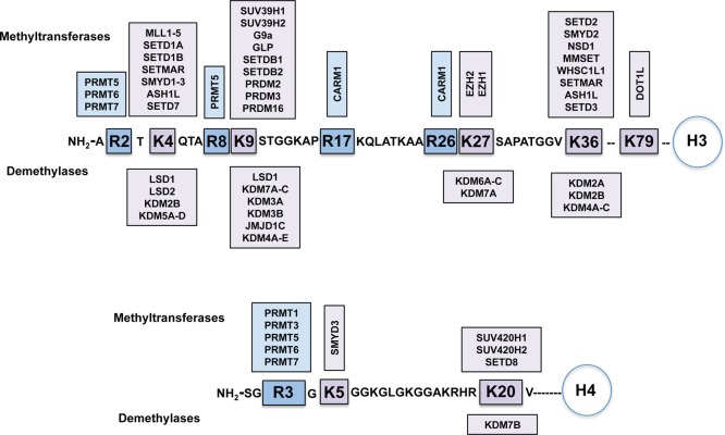
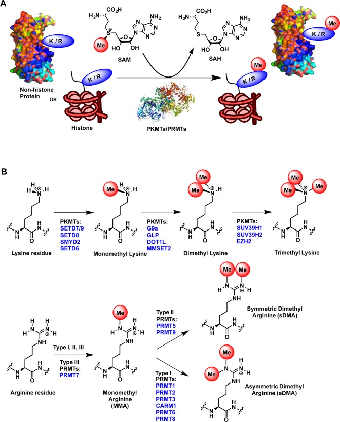
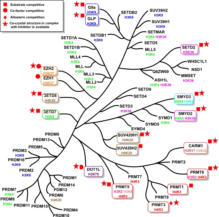
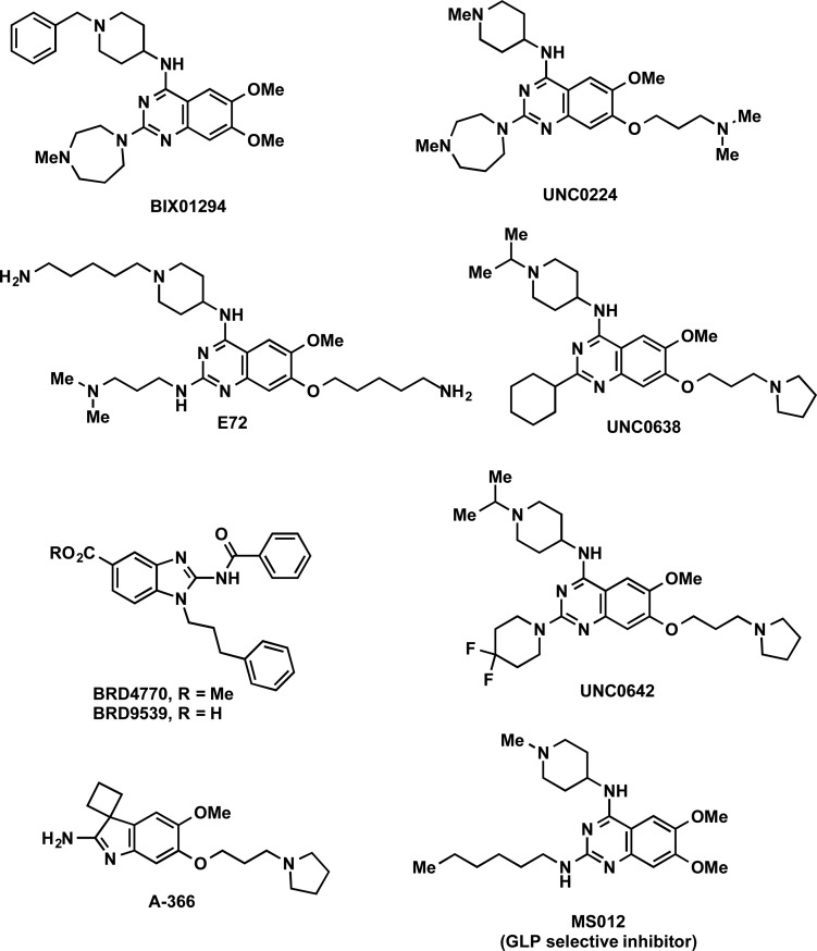
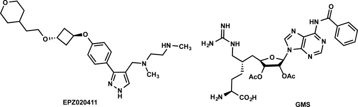
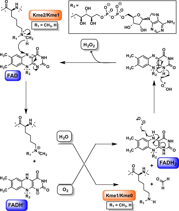
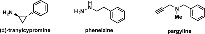
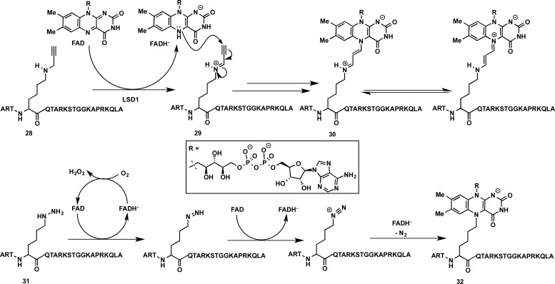
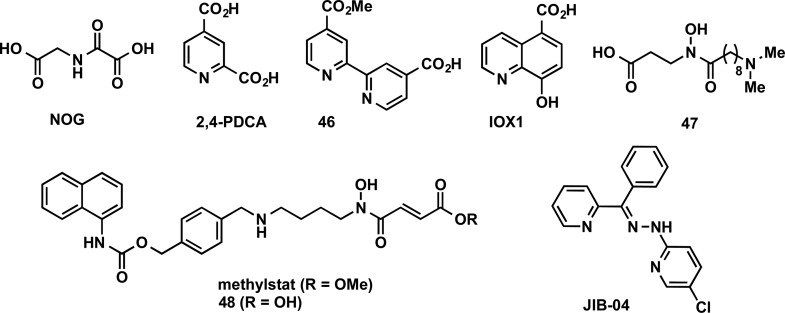
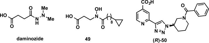
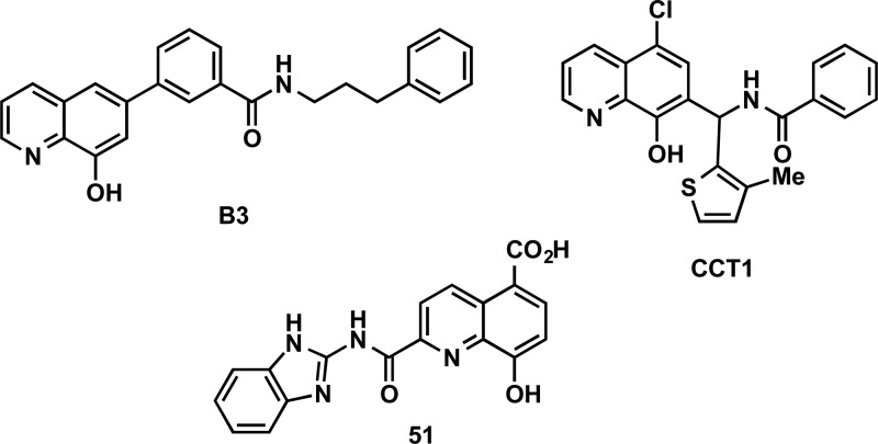
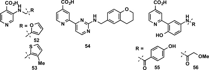
Post-translational modifications of histones by protein methyltransferases (PMTs) and histone demethylases (KDMs) play an important role in the regulation of gene expression and transcription and are implicated in cancer and many other diseases. Many of these enzymes also target various nonhistone proteins impacting numerous crucial biological pathways. Given their key biological functions and implications in human diseases, there has been a growing interest in assessing these enzymes as potential therapeutic targets. Consequently, discovering and developing inhibitors of these enzymes has become a very active and fast-growing research area over the past decade. In this review, we cover the discovery, characterization, and biological application of inhibitors of PMTs and KDMs with emphasis on key advancements in the field. We also discuss challenges, opportunities, and future directions in this emerging, exciting research field.


Conflict of interest statement

The authors declare no competing financial interest.

Figures

Figure 1
Figure 1
Known methylation and demethylation sites for histone H3 and H4 tails and corresponding protein methyltransferases and histone demethylases.
Figure 2
Figure 2
(A) Methylation of lysine (K) and arginine (R) residues of histone tails and nonhistone proteins by protein methyltransferases. (B) Methylation states of K and R residues.
Figure 3
Figure 3
Phylogenetic tree of PMTs. The PMTs with known inhibitors are indicated in boxes.
Figure 4
Figure 4
Structures of G9a/GLP inhibitors.
Figure 5
Figure 5
Co-crystal structures of UNC0224 (cyan) and UNC0638 (blue) in complex with G9a are superimposed (PDB ID: 3K5K and 3RJW, respectively). H3K9me2 peptide (magenta) overlaid for reference. Hydrogen bonds with G9a residues are represented as yellow dashed lines.
Figure 6
Figure 6
Structures of EZH2/EZH1 inhibitors.
Figure 7
Figure 7
(A) X-ray structure of Ct-PRC2 complex (PDB ID: 5CH1). (B) Co-crystal structure of PRC2 with a derivative of CPI-1205 (PDB ID: 5LS6). The key residues and interactions are indicated. SAH (blue) is overlaid for reference (PDB ID: 5HYN). Hydrogen bonds are represented as yellow dashed lines.
Figure 8
Figure 8
Structures of SETD7 inhibitors.
Figure 9
Figure 9
(A) SETD7 (light blue) in complex with (R)-PFI-2 (magenta) and SAM (yellow) (PDB ID: 4JLG) is superimposed with SETD7 structure in complex with SAH (1O9S) depicting the conformational variability of the post-SET loop. Hydrogen bonds are represented as orange dashed lines with key residues. (B) Surface representation of (R)-PFI-2-bound SETD7 highlighting an induced conformational modification of the post-SET loop.
Figure 10
Figure 10
Structures of SMYD3 inhibitors.
Figure 11
Figure 11
(A) Co-crystal structure of EPZ030456 (orange) in complex with SMYD3 (green) and SAM (yellow) (PDB ID: 5CCM). (B) Co-crystal structure of GSK2807 (orange) (PDB ID: 5HI7) in complex with SMYD3. MEKK2 (MAP3K2) peptide (magenta) and SAH (gray) is overlaid for reference (PDB ID: 5HQ8). Selected SET (green), SET-I (yellow), and post-SET (aqua) residues are indicated. Hydrogen bonds are represented as yellow dashed lines and water molecules as red spheres.
Figure 12
Figure 12
Structures of SMYD2 inhibitors.
Figure 13
Figure 13
X-ray cocrystal structure of the SMYD2-AZ-505 complex (PDB ID: 3S7B).
Figure 14
Figure 14
Structures of Sinefungin and Pr-Sinefungin (Pr-SNF).
Figure 15
Figure 15
(A) Co-crystal structure of SETD2 in complex with SAH (PDB ID: 4H12). SET domain (light orange), N-SET domain (light blue), pre-SET domain (cyan), and post-SET motif (red) are highlighted. (B) The SAH binding pocket between SET and Post-SET domains of SETD2. (C) Co-crystal structure of SETD2 in complex with Pr-SNF (PDB ID: 4FMU). (D) The key residues stabilizing the alternative configuration of the post-SET loop and interacting with Pr-SNF’s N-propyl chain.
Figure 16
Figure 16
Structures of SETD8 inhibitors.
Figure 17
Figure 17
(A) Co-crystal structure of MS2177 (orange) in complex with SETD8 (PDB ID: 5T5G). Hydrogen bonds are represented as magenta dashed lines and water molecule as red sphere. (B) Co-crystal structure of MS453 (green and cyan) in complex with SETD8 homodimer (subunits depicted in cyan and green, PDB ID: 5TH7).
Figure 18
Figure 18
Structures of additional SETD8 inhibitors.
Figure 19
Figure 19
Structure of A-196, an inhibitor of SUV420H1 and SUV420H2.
Figure 20
Figure 20
Structures of DOT1L inhibitors.
Figure 21
Figure 21
(A) Cofactor binding site of DOT1L (PDB ID: 1NW3). (B) Crystal structure of DOT1L in complex with EPZ004777 (PDB ID: 4ER3). Conformational rearrangements of DOT1L create a cavity to accommodate the t-butylphenyl group. (C) Overlay of DOT1L-SAM and DOT1L-EPZ004777 showing conformational rearrangement of substrate-binding and activation loop residues (magenta and yellow, respectively). (D) Overlay of DOT1L-EPZ004777 (PDB ID: 4ER3) and DOT1L-EPZ-5676 (PDB ID: 4HRA) complexes. Key hydrogen bonds are represented as green dashed lines between EPZ004777 (blue) and DOT1L and EPZ-5676 is depicted in orange.
Figure 22
Figure 22
Structures of recently reported DOT1L inhibitors.
Figure 23
Figure 23
Structures of type I PRMT inhibitor MS023 and reported PRMT1 inhibitors.
Figure 24
Figure 24
(A) Structures of PRMT3 inhibitors 11, 12, and 13. (B) Key interactions of 11 with PRMT3 allosteric pocket (3SMQ). (C) Structure of SGC707. (D) Co-crystal structure of PRMT3 in complex with SGC707 (PDB ID: 4RYL).
Figure 25
Figure 25
Structures of CARM1 inhibitors.
Figure 26
Figure 26
(A) Crystal structures of 14 (orange) and 15 (yellow) bound to CARM1 are superimposed (PDB ID: 2Y1W and 2Y1X). Key interactions of 14 in arginine binding channel are indicated. (B) Crystal structures of MS023 (gray) and 16 (blue) bound to PRMT6 are superimposed (PDB ID: 5E8R and 5EGS), and key interactions for 16 are shown. Hydrogen bonds are represented as magenta dashed lines and water molecule as red sphere.
Figure 27
Figure 27
Structures of PRMT5 inhibitors.
Figure 28
Figure 28
Crystal structure of EPZ015666 (orange) bound to PRMT5:MEP50 complex in the presence of SAM (yellow) (PDB ID: 4x61). Hydrogen bonds are represented as magenta dashed lines and water molecule as red spheres.
Figure 29
Figure 29
Structures of PRMT6 inhibitors.
Figure 30
Figure 30
Domain architecture of LSD1 and LSD2.
Figure 31
Figure 31
FAD-dependent enzymatic oxidation mechanism of LSDs.
Figure 32
Figure 32
Structures of mechanism-based MAO inhibitors.
Figure 33
Figure 33
Proposed structures and mechanisms of formation of FAD-(±)-tranylcypromine adduct(s).
Figure 34
Figure 34
(+)-Tranylcypromine-based inhibitors with substitution at the phenyl ring. *Absolute configuration.
Figure 35
Figure 35
(+)-Tranylcypromine-based inhibitors with substitution at both the phenyl ring and amino group. *Absolute configuration.
Figure 36
Figure 36
Structures of LSD1 inhibitors phenelzine and bizine.
Figure 37
Figure 37
Proposed structures and mechanism of formation of FAD-(±)-propargyl-lysine (30) and FAD-(±)-hydrazine-lysine derivatized peptide (32) adduct(s).
Figure 38
Figure 38
Structures of peptide-based LSD1 inhibitors.
Figure 39
Figure 39
Biguanide and bisguanidine polyamine analogues that were reported as LSD1 inhibitors.
Figure 40
Figure 40
Structures of reversible LSD1 inhibitors.
Figure 41
Figure 41
Structures of additional reversible LSD1 inhibitors.
Figure 42
Figure 42
Phylogenetic tree of JmjC KDMs.
Figure 43
Figure 43
Domain architectures of some JmjC KDMs.
Figure 44
Figure 44
Ferrous iron (Fe(II)) and 2-OG-based oxidation mechanism of JmjC KDMs.
Figure 45
Figure 45
Structures of JmjC KDMs inhibitors that contain common motifs.
Figure 46
Figure 46
Structures of KDM2/7 subfamily inhibitors.
Figure 47
Figure 47
Structures of 8HQ-containing KDM4 inhibitors.
Figure 48
Figure 48
Pyridine-4-carboxylic acid containing KDM4 inhibitors.
Figure 49
Figure 49
Structures of peptide-based KDM4 inhibitors.
Figure 50
Figure 50
Structures of other KDM4 inhibitors.
Figure 51
Figure 51
Structures of KDM5 inhibitors.
Figure 52
Figure 52
(A) KDM5-C49 (orange) binding interactions with KDM5B (PDB ID: 5A3T) and (B) GSK467 binding interactions with KDM5B (PDB ID: 5FUN). Side chains are displayed in gray, water molecules as red spheres, and the metal centers as green spheres.
Figure 53
Figure 53
Structures of other KDM5 inhibitors.
Figure 54
Figure 54
Structures of KDM6 inhibitor GSK-J1 and its prodrug GSK-J4.
Figure 55
Figure 55
Co-crystal structure of KDM6B in complex with GSK-J1 (orange). The key residues that formed the catalytic pocked are indicated in blue. Hydrogen bonds are represented as magenta dashed lines and water molecules as red spheres.
Figure 56
Figure 56
Structures of dual LSD1-JmjC KDM inhibitors.

Similar articles

Cited by

References

    1. Bernstein B. E.; Meissner A.; Lander E. S. The Mammalian Epigenome. Cell 2007, 128, 669–681. 10.1016/j.cell.2007.01.033. - DOI - PubMed
    1. Allis C. D.; Jenuwein T.; Reinberg D.. Epigenetics; Cold Spring Harbor Laboratory Press: Cold Spring Harbor, N.Y., 2007; p. 502.
    1. Gelato K. A.; Fischle W. Role of Histone Modifications in Defining Chromatin Structure and Function. Biol. Chem. 2008, 389, 353–363. 10.1515/BC.2008.048. - DOI - PubMed
    1. Rea S.; Eisenhaber F.; O’Carroll D.; Strahl B. D.; Sun Z. W.; Schmid M.; Opravil S.; Mechtler K.; Ponting C. P.; Allis C. D.; et al. Regulation of Chromatin Structure by Site-Specific Histone H3Methyltransferases. Nature 2000, 406, 593–599. 10.1038/35020506. - DOI - PubMed
    1. Shi Y.; Lan F.; Matson C.; Mulligan P.; Whetstine J. R.; Cole P. A.; Casero R. A.; Shi Y. Histone Demethylation Mediated by the Nuclear Amine Oxidase Homolog LSD1. Cell 2004, 119, 941–953. 10.1016/j.cell.2004.12.012. - DOI - PubMed

Publication types

MeSH terms