. 2017 Mar 24;18(4):693.
doi: 10.3390/ijms18040693.

Nerve Growth Factor Signaling from Membrane Microdomains to the Nucleus: Differential Regulation by Caveolins

Affiliations

Nerve Growth Factor Signaling from Membrane Microdomains to the Nucleus: Differential Regulation by Caveolins

Ambre Spencer et al. Int J Mol Sci. .

Abstract

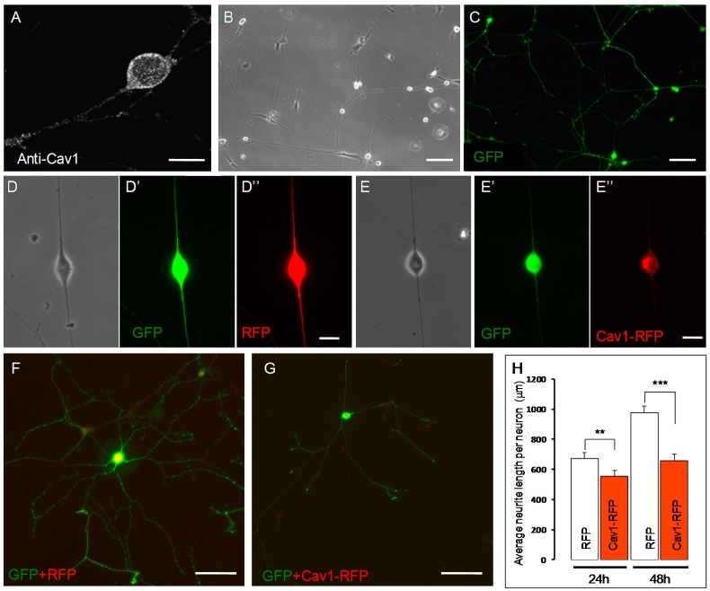
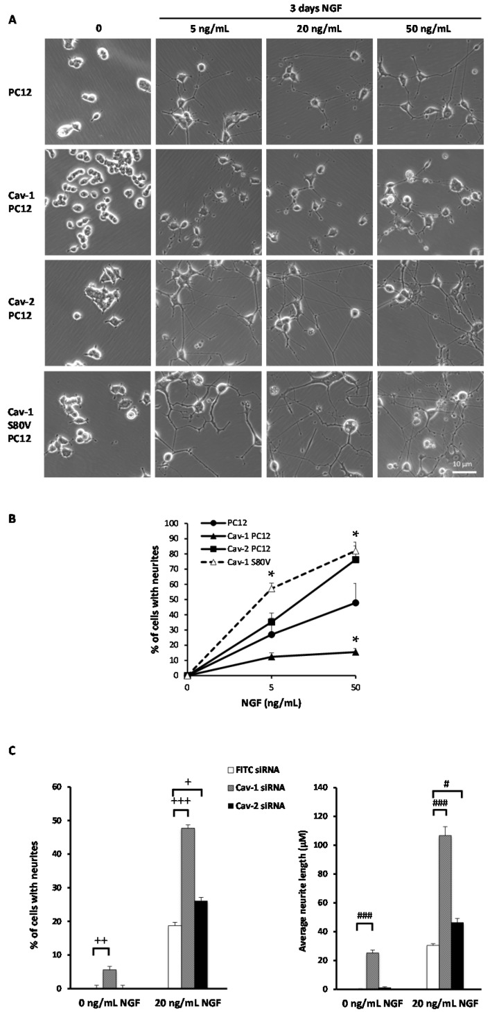
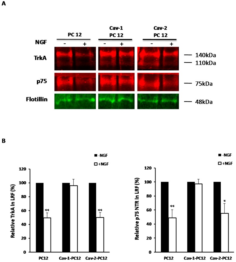
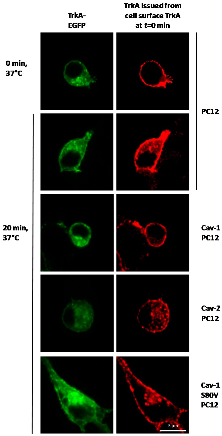
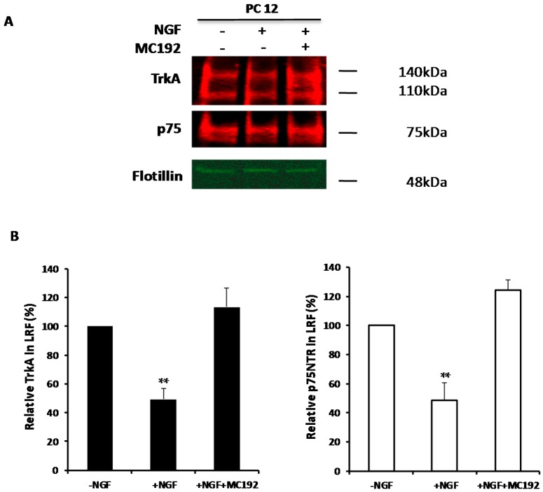
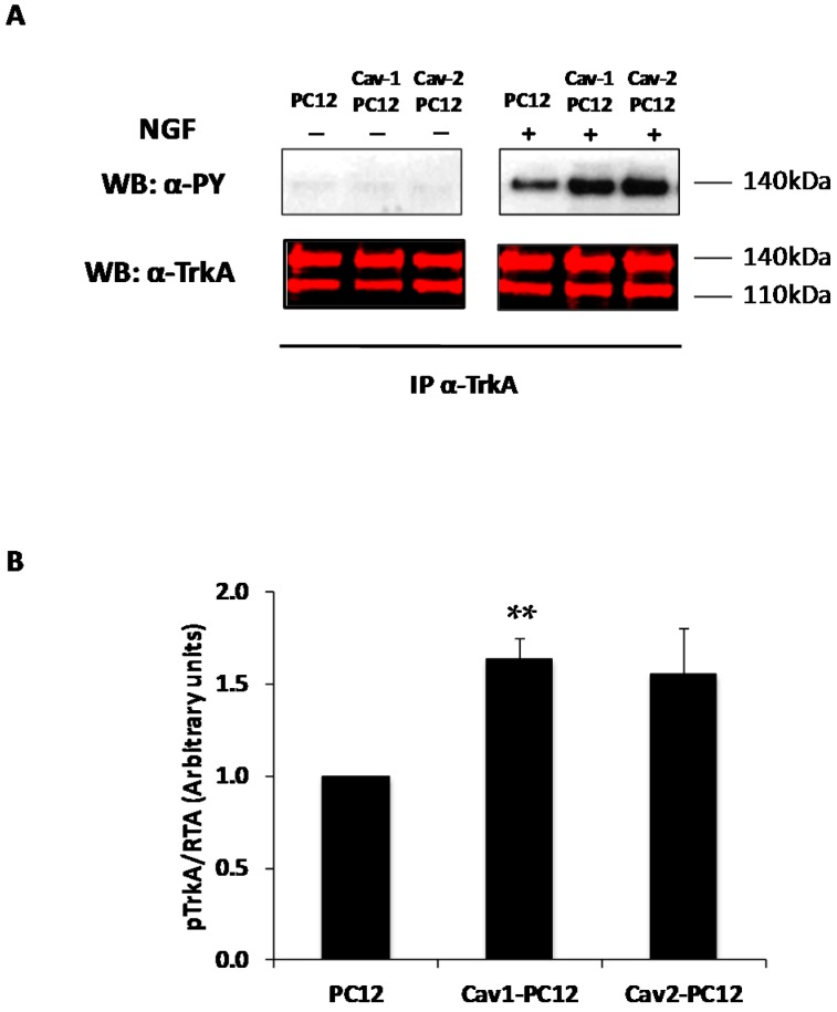
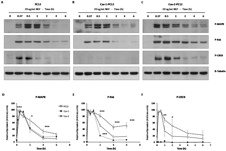
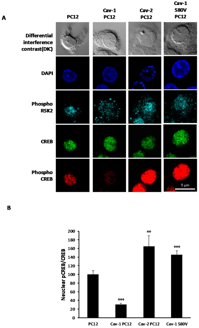
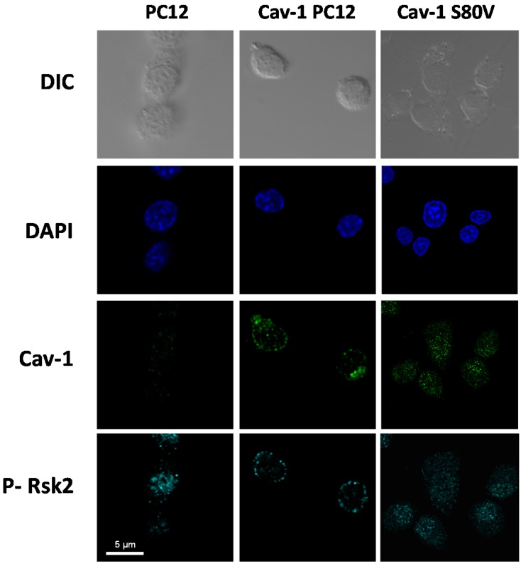
Membrane microdomains or "lipid rafts" have emerged as essential functional modules of the cell, critical for the regulation of growth factor receptor-mediated responses. Herein we describe the dichotomy between caveolin-1 and caveolin-2, structural and regulatory components of microdomains, in modulating proliferation and differentiation. Caveolin-2 potentiates while caveolin-1 inhibits nerve growth factor (NGF) signaling and subsequent cell differentiation. Caveolin-2 does not appear to impair NGF receptor trafficking but elicits prolonged and stronger activation of MAPK (mitogen-activated protein kinase), Rsk2 (ribosomal protein S6 kinase 2), and CREB (cAMP response element binding protein). In contrast, caveolin-1 does not alter initiation of the NGF signaling pathway activation; rather, it acts, at least in part, by sequestering the cognate receptors, TrkA and p75NTR, at the plasma membrane, together with the phosphorylated form of the downstream effector Rsk2, which ultimately prevents CREB phosphorylation. The non-phosphorylatable caveolin-1 serine 80 mutant (S80V), no longer inhibits TrkA trafficking or subsequent CREB phosphorylation. MC192, a monoclonal antibody towards p75NTR that does not block NGF binding, prevents exit of both NGF receptors (TrkA and p75NTR) from lipid rafts. The results presented herein underline the role of caveolin and receptor signaling complex interplay in the context of neuronal development and tumorigenesis.

Keywords: CREB; NGF; PC12; Trk; caveolin; dorsal root ganglion neurons; growth factor signaling; lipid rafts; membrane microdomains; p75NTR; trafficking.


Conflict of interest statement

The authors declare no conflict of interest.

Figures

Figure 1
Figure 1
Caveolin-1 (Cav-1) expression inhibits neurite outgrowth from mouse Dorsal root ganglia (DRG) neurons in culture. Cav-1 is detected in both the soma and the neuritic processes of E14.5 DRG neurons (A); Neon® transfection leads to efficient electroporation of E14.5 DRG neurons with little adverse effects (B,C); Neurons co-expressing GFP (Green Fluorescent Protein) and RFP (Red Fluorescent Protein) (D–D’’) or GFP and Cav-1-RFP (E–E”) can differentiate in vitro. Phase images (D and E) exemplify the morphology of GFP (D’ and E’) and RFP (D”) or Cav1-RFP (E”) expressing neurons. Nevertheless, neurons expressing Cav-1-RFP grew shorter processes than neurons expressing RFP (F,G); The length of GFP positive neurites measured and divided by the number of transfected neurons (H); Results are pooled from three sets of cultures, each culture included four mosaic fields containing >250 transfected cells. Mean ± SEM; (** p < 0.01; *** p < 0.00001). Statistical analysis was performed using the two-tail paired Student’s t-test. Scale bars represent 100 µm in B, C, F and G, and 10 µm in A, D” and E’’.
Figure 2
Figure 2
Effect of Cav-1 and Cav-2 expression on NGF-induced PC12 cell differentiation. (A) Normal PC12 cells, stably-transfected Cav-1 PC12 cells Cav-2-PC12 or Cav-1 S80V PC12 cells were plated at a density of 105 cells/well on collagen/poly-lysine coated six-well plates. Cells were maintained in DMEM (Dulbecco’s Modified Eagle’s medium) supplemented with serum for 24 h After 17 h of serum deprivation, NGF was added at increasing concentrations (0, 5, 20, 50 ng/mL NGF). After three days in culture, cells were photographed; (B) Analysis of neurite outgrowth. Quantification of the percentage of cells exhibiting neurites longer than three cell body diameters after 72 h of NGF treatment at 0, 5, and 50 ng/mL. Approximately 100 cells per condition from five independent experiments were taken into account. Values are mean percentage ± SEM. Statistical significance of the observations between PC12 cells and Cav mutant (Cav-1; Cav-2; Cav-1 S80V) PC12 cells are indicated by * p < 0.05 (unpaired, two-tail Student’s t-test). (C) Effect of siRNA towards Cav-1 and Cav-2 on neurite outgrowth. Normal PC12 cells, plated on collagen/poly-lysine coated 24-well plates (2 × 10⁴ cells per well), were transiently transfected with 30 nM siRNA against caveolin-1; caveolin-2 and FITC (fluoresceine isothiocyanate) conjugated scrambled siRNA. Cells were maintained in DMEM with 0 or 20 ng/mL NGF. After 3 days, cells were photographed and neurite outgrowth was quantified by ImageJ (Average 400 cells per condition compiled from two independent experiments. Values are mean ± SEM. Statistical significance of the observations between PC12 cells and Cav-expressing (Cav-1; Cav-2) PC12 cells are indicated by + p < 0.005; ++ p < 0.0005; +++ p < 0.00005 (% cells with neurites) and # p < 0.0005; ### p < 0.0000001 (Average neurite length) as ascertained by the unpaired, two-tail Student’s t-test).
Figure 3
Figure 3
Effect of Cav-1 and Cav-2 expression on the anti-mitogenic effect of NGF. (A) Normal PC12 cells, Cav-1 PC12 cells, Cav-2 PC12, and Cav-1 S80V PC12 cells were plated at a low density in 25 cm2 dishes containing a medium supplemented with serum. NGF 20 ng/mL was added to the medium on day 2, except for one series of dishes that was left untreated. Cell number was counted every day for six days. The experiment was performed three times, with two different clones of Cav-1 PC12 and Cav-2-PC12 cells. Cell number is normalized to cell number at day 1 (N1). Values are mean ± SEM; (B) Normal PC12 cells, Cav-1 PC12 cells, and Cav-2 PC12 cells were exposed to 20 ng/mL NGF for 0, 1, or 3 days. Proteins were extracted and resolved by SDS-PAGE and immunoblotted with CP36 monoclonal antibody directed against p21WAF1/Cip1. Equal loading was verified by reprobing the same blot with anti-Histone H1 antibody; (C) The NGF-dependent induction of p21WAF1/Cip1 was analyzed in normal PC12 cells, Cav-1 PC12 cells, and Cav-2 PC12 cells. Cells were transiently transfected with the minimal p21 promoter–Luciferase reporter (p21–Luc) and treated or not with NGF (50 ng/mL for 48 h) as described in the Materials and methods section (Mean ± SEM of three independent experiments. Statistical significance was determined using a regular two-way ANOVA test with Bonferroni post hoc tests ** p < 0.01 versus the corresponding–NGF group. ## p < 0.01, ### p < 0.001 versus the Cav-1 PC12 + NGF group).
Figure 4
Figure 4
Effect of Cav-1 and Cav-2 expression on NGF receptor exit from lipid rafts. (A) TrkA and p75NTR levels in the lipid raft fraction (LRF) before and after addition of NGF (20 ng/mL for 45 min) to cultures of normal PC12, Cav-1 PC12, and Cav-2-PC12 cells isolated, as described in the Materials and Methods. Lipid raft fractions were then subjected to Western analysis. Nitrocellulose membranes were probed with RTA, anti-p75NTR and anti-flotillin-1 antibodies. Flotillin-1 was used as a loading control. The Odyssey imaging system was used for quantitative infrared fluorescence detection of the relative amount of proteins; (B) Analysis of TrkA and p75NTR exit from lipid rafts. Level of TrkA and p75NTR in lipid rafts was normalized for flotillin-1 level for each sample. Systematic comparison of data with and without flotillin correction gave identical results. TrkA and p75NTR exit from the lipid raft fraction was then analyzed and represented as percent of the TrkA in lipid rafts compared to the amount observed in the absence of NGF, considered as 100 percent. (Mean ± SEM of three independent experiments. Statistical significance of the effect of addition of NGF vs. the absence of NGF, was ascertained using the unpaired Student’s t-Test. * p < 0.05, ** p < 0.01.)
Figure 5
Figure 5
Effect of Cav-1 and Cav-2 expression on TrkA trafficking. Internalization of TrkA was provoked by RTA addition to normal PC12, Cav-1 PC12, Cav-2 PC12, and Cav-1 S80V PC12 cells transiently expressing TrkA–EGFP chimerae (green), as described in the Materials and Methods. After fixation and permeabilization of the cell, RTA localization was determined using a secondary antibody labeled with rhodamine (red) at the indicated times. Rhodamine fluorescence indicates the location of TrkA–RTA and TrkA–EGFP–RTA complexes that were at the cell surface at the beginning of the experiment (Cell Surface TrkA at t = 0 min). Distribution of cell surface TrkA (at t = 0) in Cav-1 PC12, Cav-2-PC12, and Cav-1 S80V PC12 cells after 0 min at 37 °C is identical to that observed in PC12 cells.
Figure 6
Figure 6
Effect of MC192 on TrkA exit from lipid rafts before and after addition of NGF. (A) Lipid rafts were isolated from normal PC12 cells treated with NGF (20 ng/mL for 45 min), treated with p75NTR MC192 monoclonal antibody (8 ng/mL) for 30 min prior to and during NGF exposure (20 ng/mL for 45 min) and from untreated normal PC12 cells were isolated as described in the Materials and Methods. Lipid rafts were then subjected to Western analysis. Nitrocellulose membranes were probed with RTA and anti-flotillin-1 antibody to detect TrkA expression and flotillin-1 was used as a loading control. Revelation was achieved using quantitative infrared fluorescence detection using the Odyssey imaging system; (B) Analysis of TrkA exit from lipid rafts. Level of TrkA in the lipid rafts was normalized to the Flotillin-1 level for each sample. TrkA exit from the lipid raft fraction (LRF) was then analyzed and represented as percent of the TrkA in lipid rafts vs. TrkA in normal PC12 cells considered as 100%. (Mean ± SEM of four independent experiments. Statistical significance vs. cells in the absence of NGF, was ascertained using the unpaired, two-tail Student’s t-Test. ** p < 0.01.)
Figure 7
Figure 7
Effect of Cav-1 and Cav-2 expression on TrkA activation. Normal PC12 cells, Cav-1 PC12 cells and Cav-2 PC12 cells were left untreated or treated with NGF 20 ng/mL for 10 min after 17 h of serum deprivation. (A) One mg protein of cell lysate was immunoprecipitated with a polyclonal antibody directed against TrkA C-terminal domain (anti-Trk C-14), and immunoprecipitates were subjected to analysis by Western Blot (WB). Membrane was probed with an antibody to phosphotyrosine (α-PY) followed by a secondary antibody conjugated to horseradish peroxidase that allows chemiluminescence detection; The same blot was re-probed with RTA polyclonal antibody to TrkA followed by a secondary antibody conjugated to a fluorochrome (B), which allows infrared fluorescence detection using the Odyssey imaging system without cross-reacting with the first antibodies (Mean ± SEM from three independent experiments. Statistical significance vs. PC12 cells was ascertained using the unpaired, two-tail Student’s t-Test. ** p < 0.01).
Figure 8
Figure 8
Kinetics of activation of the MAPK pathway in PC12 cells stably-transfected with Cav-1 or Cav-2. Normal PC12 cells (A); Cav-1 PC12 cells (B) and Cav-2 PC12 cells (C) were exposed for up to 6 h to NGF 20 ng/mL. Cells were collected and proteins were extracted in lysis buffer. Protein concentrations in the lysates were determined and 40 µg were used for Western blot analysis. Blots were probed with phospho-specific antibodies to MAPK, Rsk2, and CREB. Equal loading was controlled using a β-tubulin antibody; (DF) Scans of gels were quantified using the gel analysis function of ImageJ software and analyzed as described in the Materials and Methods section; (D) P-MAPK; (E) P-Rsk; (F) P-CREB. In all, PC12 cells: Solid line with open circles; Cav-1-PC12 cells: Dotted line with filled squares; Cav-2-PC12: Dashed line with open triangles. Results are presented as the % maximal response in order to be able to compare multiple experiments. (Mean ± SEM derived from 24 gels generated in nine independent experiments performed on Cav-1 clones 3, 12, 16 and Cav-2 clones 5, 11, and 23 (see Supplementary Figure S2) having similar relative expression levels. Statistical significance was ascertained using the two-way ANOVA test with Bonferroni post hoc tests. p values are shown for results obtained with caveolin clones vs. those obtained with PC12. * p < 0.01, ** p < 0.005, *** p < 0.0005).
Figure 9
Figure 9
Effect of Cav-1, Cav-2 and Cav-1 S80V expression on TrkA effector localization. (A) Normal PC12 cells, Cav-1 PC12 cells Cav-2 PC12, and Cav-1 S80V PC12 cells were plated on collagen/poly-lysine coated coverslips and exposed to 20 ng/mL NGF for 30 min after 17 h of serum deprivation. Cells were fixed and simultaneously stained with an anti-pRsk2 antibody (cyan), an anti-CREB antibody (green) and an anti-pCREB antibody (red). Cells were mounted in mounting medium containing DAPI to visualize the nucleus; (B) Quantification of CREB phosphorylation level in the nucleus. For each single cell, quantitation of fluorescence representative of pCREB and CREB in the nucleus was assessed using ImageJ software. Level of CREB phosphorylation was evaluated by dividing pCREB fluorescence by CREB fluorescence. Results are represented as percent of CREB phosphorylation observed with CREB phosphorylation in normal PC12 cells considered as 100 percent. (Mean of 20 to 70 cells per conditions ± SEM. Statistical significance vs. PC12 cells, was ascertained using the unpaired, two-tail Student’s t-Test. ** p < 0.01; *** p < 0.001.)
Figure 10
Figure 10
Cav-1, Cav-1 S80V, and phospo-Rsk2 localization in normal, Cav-1 and Cav-1 S80V PC12 cells. Normal PC12 cells, Cav-1, and Cav-1 S80V PC12 cells were plated on collagen/poly-lysine coated coverslips and exposed to 20 ng/mL NGF for 30 min after 17 h of serum deprivation. Cells were fixed and simultaneously stained with an anti-pRsk2 antibody (cyan) and an anti-Cav-1 antibody (green).

Similar articles

Cited by

References

    1. Simons K., Toomre D. Lipid rafts and signal transduction. Nat. Rev. 2000;1:31–39. doi: 10.1038/35036052. - DOI - PubMed
    1. de Laurentiis A., Donovan L., Arcaro A. Lipid rafts and caveolae in signaling by growth factor receptors. Open Biochem. J. 2007;1:12–32. doi: 10.2174/1874091X00701010012. - DOI - PMC - PubMed
    1. Parton R.G., Simons K. The multiple faces of caveolae. Nat. Rev. 2007;8:185–194. doi: 10.1038/nrm2122. - DOI - PubMed
    1. Tang Z., Scherer P.E., Okamoto T., Song K., Chu C., Kohtz D.S., Nishimoto I., Lodish H.F., Lisanti M.P. Molecular cloning of caveolin-3, a novel member of the caveolin gene family expressed predominantly in muscle. J. Biol. Chem. 1996;271:2255–2261. - PubMed
    1. Way M., Parton R.G. M-caveolin, a muscle-specific caveolin-related protein. FEBS Lett. 1995;376:108–112. doi: 10.1016/0014-5793(95)01256-7. - DOI - PubMed

MeSH terms