Review
. 2017 Mar 24;22(4):523.
doi: 10.3390/molecules22040523.

Metal-Based PSMA Radioligands

Affiliations
Review

Metal-Based PSMA Radioligands

Eleni Gourni et al. Molecules. .

Abstract

Prostate cancer is one of the most common malignancies for which great progress has been made in identifying appropriate molecular targets that would enable efficient in vivo targeting for imaging and therapy. The type II integral membrane protein, prostate specific membrane antigen (PSMA) is overexpressed on prostate cancer cells in proportion to the stage and grade of the tumor progression, especially in androgen-independent, advanced and metastatic disease, rendering it a promising diagnostic and/or therapeutic target. From the perspective of nuclear medicine, PSMA-based radioligands may significantly impact the management of patients who suffer from prostate cancer. For that purpose, chelating-based PSMA-specific ligands have been labeled with various diagnostic and/or therapeutic radiometals for single-photon-emission tomography (SPECT), positron-emission-tomography (PET), radionuclide targeted therapy as well as intraoperative applications. This review focuses on the development and further applications of metal-based PSMA radioligands.

Keywords: PET; SPECT; intraoperative applications.; prostate specific membrane antigen (PSMA); radionuclide therapy.


Conflict of interest statement

The authors declare no conflict of interest.

Figures

Figure 1
Figure 1
The enzyme actions of PSMA. (a) Glutamic acid is released from folate polyglutamate; (b) N-Acetyl-l-aspartyl-l-glutamate (NAAG) is hydrolyzed to aspartate (NAA) and glutamate (l-Glu).
Figure 2
Figure 2
PSMA inhibitors. 2-PMPA (1) is the first potent PSMA inhibitor. The three groups of PSMA inhibitors include: thiols 2 and 3, phosphonates 4 and ureas 5 and 6.
Figure 3
Figure 3
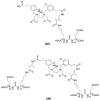
Chemical structures of the 99mTc-labeled GPI (7) GPI monomer (8), GPI dimer (9) and GPI trimer (10) [72,73].
Figure 4
Figure 4
Chemical structure of 99mTc(CO)3-DTPA-CTT-54 (11) [74].
Figure 5
Figure 5
Chemical structure of 64Cu-ABN-1 (12) [75].
Figure 6
Figure 6
PSMA-inhibitors labeled via the ([99mTcO]3+) (1320), ([99mTc(CO)3]+) (2124) and (99mTc-hynic) (25) core [98,99].
Figure 7
Figure 7
99mTc-labeled PSMA inhibitors (2631) [100].
Figure 8
Figure 8
Chemical structure of 99mTc-MAS3 (32)/mas3 (33)-y-nal-k-Sub-KuE (MAS3/mas3-D-Tyr-D-2-Nal-D-Lys-suberoyl-Lys-urea-Glu) [101].
Figure 9
Figure 9
Chemical structure of 99mTc-TMCE (34) [102].
Figure 10
Figure 10
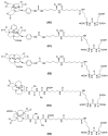
Chemical structures of 99mTc-MIP-1428 (35), 99mTc-MIP-1405 (36), 99mTc-MIP-1404 (37) and 99mTc-MIP-1427 (38) [103].
Figure 11
Figure 11
Chemical structures of the monomer 68Ga-PSMA-11 (39) and the dimer 68Ga-PSMA-10 (40) [106,130].
Figure 12
Figure 12
Chemical structures of the two DOTA- (41 and 42) and the NOTA-conjugated (43) PSMA inhibitors labeled with 68Ga [131,132].
Figure 13
Figure 13
Chemical structure of 68Ga-PSMA-617 (44) [133].
Figure 14
Figure 14
Chemical structures of the three generations (DOTAGA-FFK(Sub-KuE) (45), DOTAGA-ffk(Sub-KuE) (46) and of DOTAGA-(I-y)fk(Sub-KuE) (47)) PSMA inhibitors labeled with 68Ga [134,135].
Figure 15
Figure 15
Chemical structure of 68Ga-(R)-NODAGA-Phe-Phe-d-Lys(suberoyl)-Lys-urea-Glu (68Ga-CC34) (48) [136].
Figure 16
Figure 16
Chemical structure of 68Ga-CHX-A″-DTPA-DUPA-Pep (49) [137].
Figure 17
Figure 17
Chemical structures of the PSMA-based inhibitors (5054) labeled with 64Cu [139].
Figure 18
Figure 18
Tumor uptake of 99mTc-MIP-1404 (A) or 99mTc-MIP-1405 (B) at 4 h in patient with metastatic prostate cancer, compared with that of standard bone scan (C). Images also show uptake of radiotracer in normal parotid and salivary glands. Ant = anterior; Post = posterior (Reprinted with permission of [143]).
Figure 19
Figure 19
68Ga-PSMA-11 PET/CT image of a patient with locally recurrent prostate cancer (PSA 3.7 ng/mL) after radical prostatectomy (SUVmax 12.4) who received 140 MBq of the 68Ga-labeled tracer molecule and was scanned at 1 h p.i.; (a) CT image; (b) PET image; (c) PET/CT fusion image; (d) MIP (Reprinted with permission of [145]).
Figure 20
Figure 20
(AC) 68Ga-PSMA-617 PET/CT of a patient at 1 h after injection. Red arrows point to a bone metastasis with SUVmax of 21.7 at 1 h and 32.6 at 3 h after injection. (A) Low dose CT; (B) Fusion of PET and CT; (C) MIP of PET/CT. MIP = maximum-intensity projection (Reprinted with permission of [146]).
Figure 21
Figure 21
70-year-old patient with PSMA-avid lymph node metastases on 68Ga-PSMA PET/CT before therapy (A) and on 177Lu-PSMA I&T scintigraphy after first PSMA radionuclide therapy (B); with remarkable reduction in uptake after second PSMA RLT (C). Results were consistent with excellent therapy response projection (Reprinted with permission of [153]).

Similar articles

Cited by

References

    1. Hricak H., Choyke P.L., Eberhardt S.C., Leibel S.A., Scardino P.T. Imaging prostate cancer: A multidisciplinary perspective. Radiology. 2007;243:28–53. doi: 10.1148/radiol.2431030580. - DOI - PubMed
    1. Beer A.J., Eiber M., Souvatzoglou M., Schwaiger M., Krause B.J. Radionuclide and hybrid imaging of recurrent prostate cancer. Lancet Oncol. 2011;12:181–191. doi: 10.1016/S1470-2045(10)70103-0. - DOI - PubMed
    1. Fani M., Maecke H.R. Radiopharmaceuticals development of radiolabelled peptides. Eur. J Nucl. Med. Mol. Imaging. 2012;1:S11–S30. doi: 10.1007/s00259-011-2001-z. - DOI - PubMed
    1. Schottelius M., Wester H.J. Molecular imaging targeting peptide receptors. Methods. 2009;48:161–177. doi: 10.1016/j.ymeth.2009.03.012. - DOI - PubMed
    1. Chang S.S. Overview of prostate-specific membrane antigen. Rev. Urol. 2004;6(Suppl. 10):S13–S18. - PMC - PubMed