

Type IV Secretion and Signal Transduction of Helicobacter pylori CagA through Interactions with Host Cell Receptors
- PMID: 28338646
- PMCID: PMC5408189
- DOI: 10.3390/toxins9040115
Type IV Secretion and Signal Transduction of Helicobacter pylori CagA through Interactions with Host Cell Receptors
Abstract
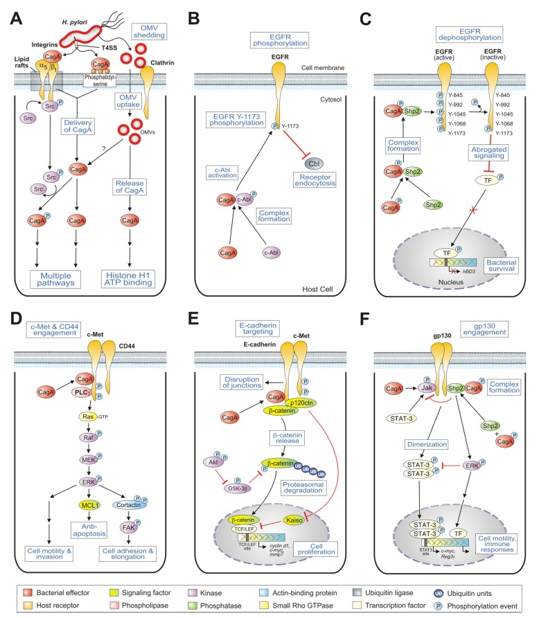
Helicobacter pylori is a highly successful human bacterium, which is exceptionally equipped to persistently inhabit the human stomach. Colonization by this pathogen is associated with gastric disorders ranging from chronic gastritis and peptic ulcers to cancer. Highly virulent H. pylori strains express the well-established adhesins BabA/B, SabA, AlpA/B, OipA, and HopQ, and a type IV secretion system (T4SS) encoded by the cag pathogenicity island (PAI). The adhesins ascertain intimate bacterial contact to gastric epithelial cells, while the T4SS represents an extracellular pilus-like structure for the translocation of the effector protein CagA. Numerous T4SS components including CagI, CagL, CagY, and CagA have been shown to target the integrin-β₁ receptor followed by translocation of CagA across the host cell membrane. The interaction of CagA with membrane-anchored phosphatidylserine and CagA-containing outer membrane vesicles may also play a role in the delivery process. Translocated CagA undergoes tyrosine phosphorylation in C-terminal EPIYA-repeat motifs by oncogenic Src and Abl kinases. CagA then interacts with an array of host signaling proteins followed by their activation or inactivation in phosphorylation-dependent and phosphorylation-independent fashions. We now count about 25 host cell binding partners of intracellular CagA, which represent the highest quantity of all currently known virulence-associated effector proteins in the microbial world. Here we review the research progress in characterizing interactions of CagA with multiple host cell receptors in the gastric epithelium, including integrin-β₁, EGFR, c-Met, CD44, E-cadherin, and gp130. The contribution of these interactions to H. pylori colonization, signal transduction, and gastric pathogenesis is discussed.
Keywords: Janus kinase; STAT3; cortactin; glycogen synthase kinase 3beta GSK3β; p120 catenin; phosphatase Shp-2; phosphatidylinositol 3-kinase PI3K; portioning kinase Par1b; protein kinase Akt; β-catenin.
Conflict of interest statement
The authors declare no conflict of interest.
Figures
References
-
- Huang J.Y., Sweeney E.G., Sigal M., Zhang H.C., Remington S.J., Cantrell M.A., Kuo C.J., Guillemin K., Amieva M.R. Chemodetection and Destruction of Host Urea Allows Helicobacter pylori to Locate the Epithelium. Cell Host Microbe. 2015;18:147–156. doi: 10.1016/j.chom.2015.07.002. - DOI - PMC - PubMed
Publication types
MeSH terms
Substances
LinkOut - more resources
Full Text Sources
Other Literature Sources
Research Materials
Miscellaneous
