. 2017 Mar 1;68(7):1425-1440.
doi: 10.1093/jxb/erx024.

Subcellular localization of Arabidopsis arogenate dehydratases suggests novel and non-enzymatic roles

Affiliations

Subcellular localization of Arabidopsis arogenate dehydratases suggests novel and non-enzymatic roles

Crystal D Bross et al. J Exp Bot. .

Abstract

Arogenate dehydratases (ADTs) catalyze the final step in phenylalanine biosynthesis in plants. The Arabidopsis thaliana genome encodes a family of six ADTs capable of decarboxylating/dehydrating arogenate into phenylalanine. Using cyan fluorescent protein (CFP)-tagged proteins, the subcellular localization patterns of all six A. thaliana ADTs were investigated in intact Nicotiana benthamiana and A. thaliana leaf cells. We show that A. thaliana ADTs localize to stroma and stromules (stroma-filled tubules) of chloroplasts. This localization pattern is consistent with the enzymatic function of ADTs as many enzymes required for amino acid biosynthesis are primarily localized to chloroplasts, and stromules are thought to increase metabolite transport from chloroplasts to other cellular compartments. Furthermore, we provide evidence that ADTs have additional, non-enzymatic roles. ADT2 localizes in a ring around the equatorial plane of chloroplasts or to a chloroplast pole, which suggests that ADT2 is a component of the chloroplast division machinery. In addition to chloroplasts, ADT5 was also found in nuclei, again suggesting a non-enzymatic role for ADT5. We also show evidence that ADT5 is transported to the nucleus via stromules. We propose that ADT2 and ADT5 are moonlighting proteins that play an enzymatic role in phenylalanine biosynthesis and a second role in chloroplast division or transcriptional regulation, respectively.

Keywords: Arogenate dehydratase; chloroplast division; moonlighting proteins; nuclear localization; phenylalanine biosynthesis; stromules..


Figures

Fig. 1.
Fig. 1.
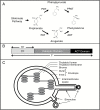
Phenylalanine synthesis, arogenate dehydratases, and stromules. (A) Phenylalanine can be synthesized in plants using either the prephenate (top) or the arogenate (bottom) pathway (Cho et al., 2007; Maeda and Dudareva, 2012). Prephenate is either decarboxylated/dehydrated to phenylpyruvate (PP) by a prephenate dehydratase (PDT) and PP is then transaminated by a phenylpyruvate aminotransferase (PPAT) to phenylalanine. Alternatively the two enzymatic steps are reversed, whereby prephenate is transaminated to arogenate by a prephenate aminotransferase (PAT) and arogenate is then decarboxylated/dehydrated to phenylalanine by an arogenate dehydratase (ADT). (B) A. thaliana ADT constructs were cloned in different lengths. The full-length (F) sequence represents the entire ADT ORF while an N-terminal construct only includes the transit peptide (TP). (C) Schematic diagram of a chloroplast showing the formation of stromules. Stromules are stroma-filled protrusions of the outer and inner membrane from chloroplasts. They can differ in length, forming long thread-like extensions or globular structures.
Fig. 2.
Fig. 2.
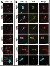
Subcellular localization of ADT–FP fusion proteins and co-localization with TP-ssRuBisCO–YFP. (A) ADT–CFP subcellular localization patterns. ADT1–ADT5 localized to stroma and to areas seemingly close to the chloroplast just outside of the autofluorescence signal generated by chlorophyll. They often appear either in thread-like structures (e.g. the arrow in ADT2) or globular structures (e.g. the arrows in ADT4). The ADT6–CFP pattern is distinctly different, showing a cytosolic distribution. Images were taken at a lower magnification to allow observation of the CFP signal relative to several chloroplasts. (B) Close-ups of ADT–CFP subcellular localization patterns in relation to TP-ssRuBisCO–YFP. In contrast to the chlorophyll autofluorescence, the TP-ssRuBisCO–YFP is a stroma-specific marker that visualizes all stroma-filled areas within the chloroplast including stromules. ADT1–ADT5 are found within the main body of chloroplast and in stromules, while ADT6 is found within the cytosol and does not co-localize with TP-ssRuBisCO–YFP.
Fig. 3.
Fig. 3.
Localization of ADTs to the chloroplast is dependent on the transit peptide sequences. To test if the transit peptide sequences are sufficient for the transport of ADTs to the chloroplast, the first 99 amino acids of ADT2 (TP-ADT2–CFP) were expressed transiently in N. benthamiana leaves.
Fig. 4.
Fig. 4.
ADT2 forms structures consistent with chloroplast division rings. (A) In addition to being expressed within chloroplasts and stromules, ADT2 was also found to accumulate in places consistent with chloroplast division rings. The top panel shows ADT2 forming rings at the equatorial plate of the chloroplast. On occasion, ADT2 was found in the constriction zone of chloroplasts (two middle panels). In these cases, the chloroplasts have a distinct dumb-bell shape and the degree of indentation depends on how far the division process has proceeded. In addition, ADT2 accumulated in a spindle-like shape that tapers at chloroplast poles (bottom panel). This fusiform ADT2 accumulation was only found at one pole of the chloroplast and is distinct from a stromule pattern shown in Fig. 2. (B) Schematic of chloroplast division stages: from top to bottom, positioning of chloroplast division rings; slightly constricted chloroplast just prior to division; two daughter chloroplast following division. Analogous to the fluorescent images, chloroplasts are shown in red and the position of ring proteins in blue (adapted from Miyagishima, 2011).
Fig. 5.
Fig. 5.
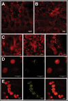
Chloroplast morphology and FtsZ2–YFP localization is affected by a point mutation in ADT2. (A) Chloroplasts in wild-type A. thaliana Col-0. (B) Chloroplasts in adt2-1D A. thaliana mutants. (C) Close-ups of chloroplasts observed in adt2-1D to show the heterogeneity in shape and size. (D) Transiently expressed FtsZ–YFP in wild-type Col-0 localizes as expected to a single ring at the equatorial plane. (E) In contrast, FtsZ2–YFP localizes as long spiralling filaments within adt2-1D chloroplasts. (D, E) Images of chlorophyll fluorescence (left) and FtsZ2–YFP (middle) are shown separately and merged (right).
Fig. 6.
Fig. 6.
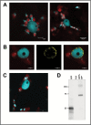
ADT5 is found in the nucleus. ADT5–CFP proteins are unique as they are the only full-length ADT proteins that were found in the nucleus. (A) Nuclei show a close association with chloroplasts (left) or with stromules of chloroplasts (right). Both images show ADT5–CFP within nuclei. (B) Co-localization of ADT5–CFP with NUP1–YFP. To determine if ADT5–CFP localizes to the nucleus, it was co-expressed with NUP1–YFP in N. benthamiana. Images of chlorophyll fluorescence and ADT5–CFP are shown merged (left). NUP1–YFP is shown alone (middle) and merged with ADT5–CFP and chlorophyll fluorescence (right). NUP1–YFP localizes to the nuclear membrane and surrounds ADT5–CFP, confirming that it localizes to the nucleus. (C) ADT5–CFP transiently expressed with its native ADT5 promoter also localizes to the nucleus. (D) Western blot of ADT5–CFP (calculated size 73.9 kDa) expressed with its native promoter and visualized with a GFP antibody is detected at its expected size. As negative controls, proteins isolated from leaves transformed with GFP (25 kDa) and p19 are shown. Total soluble protein was isolated from transiently transformed leaves, and 10 μg of total soluble protein was size separated by 10% SDS–PAGE. Sizes of the protein ladder are given in kDa.
Fig. 7.
Fig. 7.
The presence of ADT5 in the nucleus is affected by the ability to form stromules. To determine if nuclear localization of ADT5 is dependent on stromules, plants were co-infiltrated with TP-ADT2–CFP (A and B) as a control or ADT5–CFP (C) and an empty vector (dark gray), dominant negative myosin XI-2 (dnMyoXI-2; light gray) and myosin XI-K (dnMyoXI-K/GTD; white), respectively. (A) Percentage of chloroplasts having stromules. Chloroplasts were analyzed if they contained any visible TP-ADT2–CFP fluorescence and were determined to have a stromule if the projection was longer than 1 μm. In total 554, 395, and 579 chloroplasts were analyzed from plants transformed with an empty vector, dnMyoXI-2, and dnMyoXI-K/GTD, respectively. (B) Average length of stromules. A total of 166, 93, and 91 stromules were measured from plants transformed with an empty vector, dnMyoXI-2, and dnMyoXI-K/GTD, respectively. (C) Nuclear localization of ADT5–CFP. Cells were analyzed for CFP fluorescence in the nucleus only if any ADT5–CFP fluorescence was detectable. A total of 131, 190, and 358 cells were analyzed from plants transformed with an empty vector, dnMyoXI-2, and dnMyoXI-K/GTD, respectively. Each experiment was performed on three independent occasions. Significant differences (P<0.001) as determined by a one-way ANOVA (multiple comparisons) are indicated by different letters. Averages ± SE of the mean are plotted.
Fig. 8.
Fig. 8.
ADT localization to the stroma and stromules, the chloroplast equatorial plane, and the nucleus can also be detected in A. thaliana. To test if the ADT patterns determined in N. benthamiana reflect expression in A. thaliana, all six ADT–CFP fusion proteins were transiently expressed in A. thaliana Col-0. All images show a merge of chlorophyll and CFP fluorescence. (A) ADT1–CFP through ADT5–CFP localize to stroma and structures resembling stromules of varying shapes and lengths, with varying levels of fluorescence in the stroma. ADT6–CFP localizes outside of chloroplasts in the cytosol. (B) Chloroplast division patterns for ADT2–CFP. (C) Nuclear localization of ADT5–CFP.

Similar articles

Cited by

References

    1. Agati G, Biricolti S, Guidi L, Ferrini F, Fini A, Tattini M. 2011. The biosynthesis of flavonoids is enhanced similarly by UV radiation and root zone salinity in L. vulgare leaves. Journal of Plant Physiology 168, 204–212. - PubMed
    1. Avisar D, Prokhnevsky AI, Makarova KS, Koonin EV, Dolja VV. 2008. Myosin XI-K is required for rapid trafficking of Golgi stacks, peroxisomes, and mitochondria in leaf cells of Nicotiana benthamiana. Plant Physiology 146, 1098–1108. - PMC - PubMed
    1. Bertani G. 2004. Lysogeny at mid-twentieth century: P1, P2, and other experimental systems. Journal of Bacteriology 186, 595–600. - PMC - PubMed
    1. Bross CD, Corea ORA, Kaldis A, Menassa R, Bernards MA, Kohalmi SE. 2011. Complementation of the pha2 yeast mutant suggests functional differences for arogenate dehydratases from Arabidopsis thaliana. Plant Physiology and Biochemistry 49, 882–890. - PubMed
    1. Cho MH, Corea ORA, Yang H, et al. 2007. Phenylalanine biosynthesis in Arabidopsis thaliana. Identification and characterization of arogenate dehydratases. Journal of Biological Chemistry 282, 30827–30835. - PubMed

Publication types

MeSH terms