. 2017 Aug 1;19(8):1068-1078.
doi: 10.1093/neuonc/now299.

The transcription factor Olig2 is important for the biology of diffuse intrinsic pontine gliomas

Affiliations

The transcription factor Olig2 is important for the biology of diffuse intrinsic pontine gliomas

Jane L Anderson et al. Neuro Oncol. .

Abstract

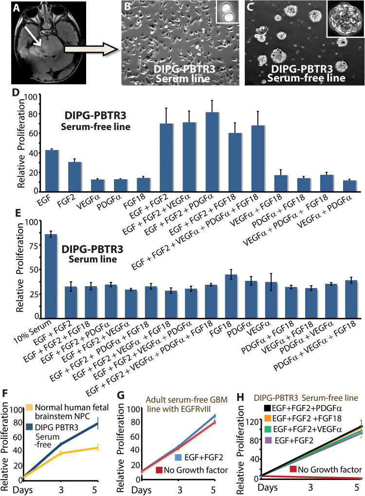
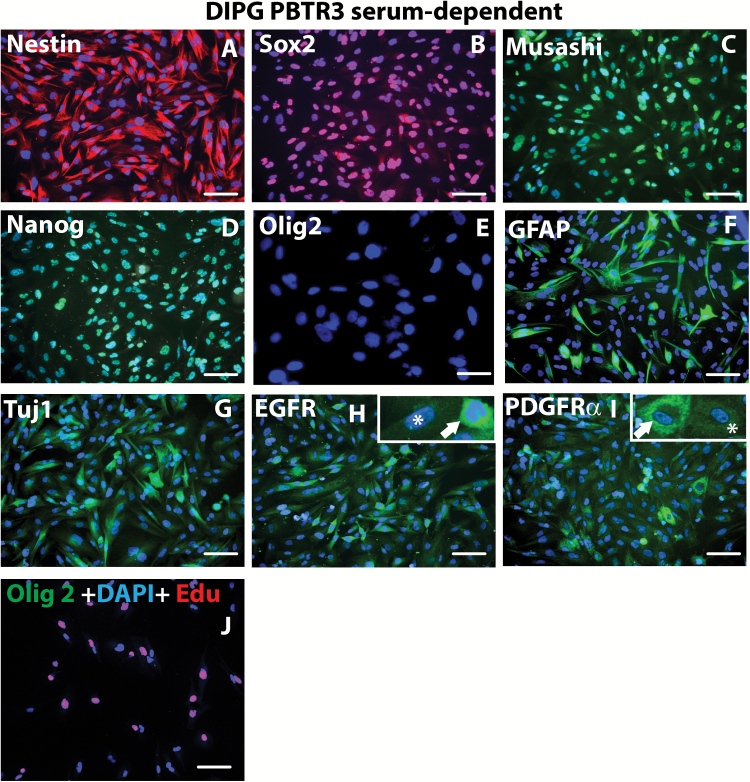
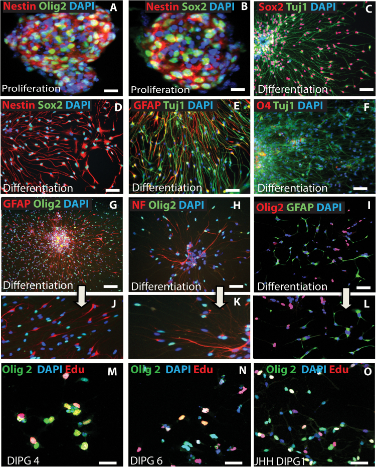
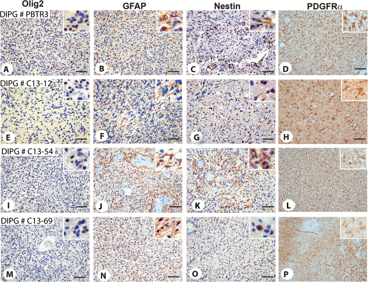
Background: Diffuse intrinsic pontine glioma (DIPG) is a high-grade brainstem glioma of children with dismal prognosis. There is no single unifying model about the cell of origin of DIPGs. Proliferating cells in the developing human and mouse pons, the site of DIPGs, express neural stem/progenitor cell (NPC) markers, including Sox2, nestin, vimentin, Olig2, and glial fibrillary acidic protein, in an overlapping and non-overlapping manner, suggesting progenitor cell heterogeneity in the pons. It is thought that during a restricted window of postnatal pons development, a differentiation block caused by genetic/epigenetic changes leads to unrestrained progenitor proliferation and DIPG development. Nearly 80% of DIPGs harbor a mutation in the H3F3A or the related HIST1H3B gene. Supporting the impaired differentiation model, NPCs derived from human induced pluripotent stem cells expressing the H3F3A mutation showed complete differentiation block. However, the mechanisms regulating an altered differentiation program in DIPG are unknown.

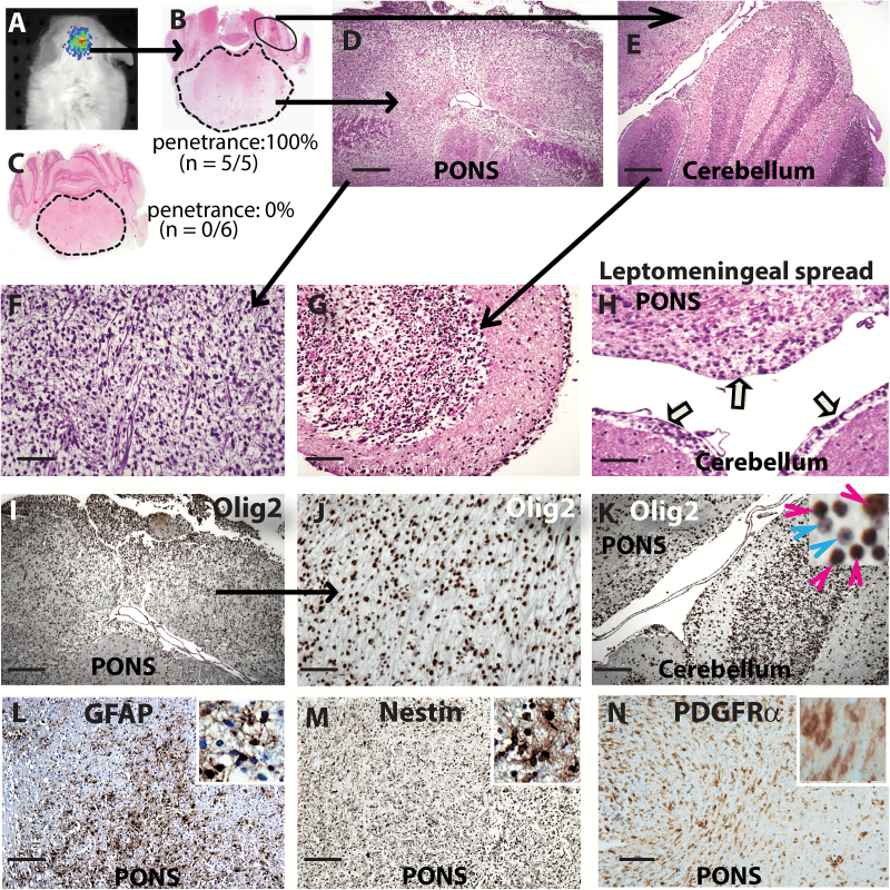
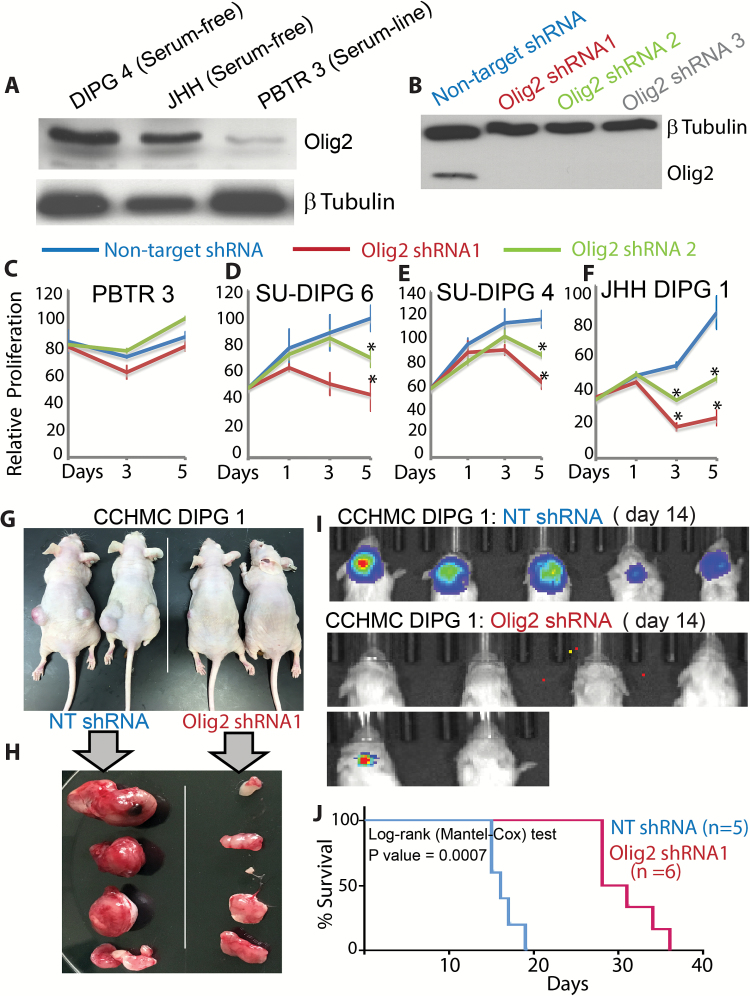
Methods: We established syngeneic serum-dependent and independent primary DIPG lines, performed molecular characterization of DIPG lines in vitro and in an orthotopic xenograft model, and used small hairpin RNA to examine Olig2 function in DIPG.

Results: The transcription factor Olig2 is highly expressed in 70%-80% of DIPGs. Here we report that Olig2 expression and DIPG differentiation are mutually exclusive events in vitro, and only DIPG cells that retained Olig2 in vitro formed robust Olig2-positive brainstem glioma with 100% penetrance in a xenograft model.

Conclusion: Our results indicate Olig2 as an onco-requisite factor in DIPG and propose investigation of Olig2 target genes as novel candidates in DIPG therapy.

Keywords: DIPG; Olig2; differentiation.


Figures

Fig. 1
Fig. 1
Establishment of serum-free and serum-dependent DIPG lines. (A) Axial MRI scan showing location of the DIPG in the patient # PBTR3. The tumor is shown by the arrow. Bright field images of the serum-dependent line (B) and serum-free sphere line (C) established from autopsy tissue. Proliferation of serum-free line (D) and serum line (E) in the presence of combination of growth factors or serum. (F) Relative proliferation of serum-free DIPG line and normal human fetal brainstem neural progenitor cells (NPC) grown in identical conditions. (G) Proliferation of an adult GBM serum-free line in the presence or absence of growth factors. (H) Proliferation of the serum-free DIPG line in the presence or absence of growth factors.
Fig. 2
Fig. 2
Immunophenotyping of PBTR3 serum line. Confocal immunofluorescence microscopy images (magnification 20×) of PBTR3 serum line showing marker expression: (A‒E) showing expression of stem cell/proliferation markers—nestin, Sox2, Musashi, Nanog, and Olig2. Note: the serum line expresses the typical stem cell/NPC markers except Olig2. Expression of the astrocytic marker (F) and the early neuronal marker Tuj1 (G) and that of 2 receptor tyrosine kinases, EGFR (H) and PDGFRα, (I) are shown. The arrows and asterisks in (H) and (I) shows heterogeneous expression of the receptor tyrosine kinases. (J) DNA synthesis (EdU incorporation) and Olig2 expression in reduced serum (5.0 serum %) condition. Nuclei were stained with 4′,6′-diamidino-2-phenylindole. Scale bars 2 µm.
Fig. 3
Fig. 3
Mutually exclusive expression of Olig2 and differentiation markers during DIPG differentiation in vitro. (A‒H & J, K = PBTR3 serum-free; I, L = JHH-DIPG). Confocal immunofluorescence microscopy images showing marker expression during DIPG proliferation and differentiation: (A, B) expression of nestin and Olig2 in DIPG spheres in proliferation condition; (C‒L) marker expression during differentiation condition in the absence of growth factors. Note: DIPG cells under this condition continue to express stem cell markers Sox2 (C), nestin (D), and Olig2 (G–I); a fraction of cells also express the astrocytic marker GFAP and the early neuronal marker Tuj1 (E), and the mature neuronal marker neurofilament (H); very few cells expressed the oligodendrocyte marker O4 (F). Magnified images of G, H, and I showing mutual exclusivity between Olig2 and GFAP (J, L) and Olig2 and NF (K) in PBTR3 and JHH DIPG1. (M‒O) Edu incorporation assay, showing DNA synthesis in Olig2+ cells under differentiation condition. Nuclei were stained with 4′,6′-diamidino-2-phenylindole. Scale bars 2 µm (A‒I); 100 µm (M‒O).
Fig. 4
Fig. 4
Establishment of xenograft from PBTR3 serum-free line. (A) Imaging of luciferase-positive DIPG xenograft in the brainstem of NSG mice. (B, C) Hematoxylin and eosin (H&E) staining showing brainstem and cerebellum (magnification 1.6×) of mice injected with serum-free PBTR3 line (B) or isogenic serum line (C). Note: enlarged pons in (B) but not in (C). (D, E) Magnified images of pons and cerebellum of mice injected with the serum-free line showing high cellularity in both structures. (F, G) Magnified images of pons and cerebellum showing diffuse nature of the tumor in the pons (F) and infiltration of tumor cells in the cerebellar folia (G). (H) H&E image showing tumor cell spread across the meninges akin to leptomeningeal disease (arrows). (I‒K) IHC showing Olig2 positivity of nearly all tumor cells in the pons (I, J) and the cerebellum (K; inset magnification 63×) which shows infiltration of Olig2+ DIPG cells (red arrowhead) among granule cell neurons (blue arrowhead). (L, M, N) GFAP, nestin, and PDGFRα immunoreactivity, respectively. Scale bars 500 μm (D, E, I, K‒N); 200 μm (F, G,H, J).
Fig. 5
Fig. 5
Immunophenotyping of original PBTR3 and other DIPGs. IHC of original PBTR3 DIPG and 3 other DIPGs showing expression of Olig2 (A, E, I, M), GFAP (B, F, J, N), nestin (C, G, K, O), and PDGFRα (D, H, L, P). Magnifications 20× (A‒P); all insets equal area cropped 40×. Note that 3 out of 4 tumors express high Olig2 (A, D, G); GFAP was high in 2 tumors, DIPG # C13-54 (H) and C13-69 (K); only a few GFAP+ tumor cells were observed in PBTR3 and DIPG #C13-12 (B, E, inset); nestin expression was generally low except DIPG #C13-54 (I), while all tumors expressed high levels of PDGFRα. In the original PBTR3, nestin expression was primarily restricted to endothelial cells (C; inset in C shows a few nestin+ tumor cells). Scale bars 0.005 mm.
Fig. 6
Fig. 6
Olig2 is required for DIPG proliferation. (A) Western blot showing Olig2 expression in serum-free DIPG4, JHH lines, and serum-dependent PBTR3 DIPG line. (B) Western blot showing Olig2 knockdown by Olig2 shRNA but not by control nontarget shRNA in DIPG4. (C‒F) Relative proliferation of PBTR3 serum line and serum-free SU-DIPG6, SU-DIPG4, and JHH-DIPG1 lines expressing nontarget or Olig2 shRNA. *P ≤ .005. Photomicrographs showing flank tumor formation in situ (G) or post resection (H) in Nu/Nu mice by CCHMC-DIPG1 serum-free line expressing nontarget shRNA or Olig2 shRNA. (I) NSG mice were imaged using the IVIS (Xenogen) system to monitor tumor growth at indicated days. (J) Kaplan‒Meier survival data of CCHMC-DIPG1 serum-free line expressing NT or Olig2 shRNA. *P = .0007.

References

    1. Ostrom QT, Gittleman H, Liao P. CBTRUS statistical report: primary brain and central nervous system tumors diagnosed in the United States 2007–2011. Neuro Oncol. 2014;16(4):1–63. - PMC - PubMed
    1. Warren KE. Diffuse intrinsic pontine glioma: poised for progress. Front Oncol. 2012;2:205. - PMC - PubMed
    1. Caretti V, Bugiani M, Freret M, et al. Subventricular spread of diffuse intrinsic pontine glioma. Acta Neuropathol. 2014;128(4):605–607. - PMC - PubMed
    1. Buczkowicz P, Bartels U, Bouffet E, Becher O, Hawkins C. Histopathological spectrum of paediatric diffuse intrinsic pontine glioma: diagnostic and therapeutic implications. Acta Neuropathol. 2014;128(4):573–581. - PMC - PubMed
    1. Fisher PG, Breiter SN, Carson BS, et al. A clinicopathologic reappraisal of brain stem tumor classification. Identification of pilocystic astrocytoma and fibrillary astrocytoma as distinct entities. Cancer. 2000;89(7):1569–1576. - PubMed

Substances