. 2017 Apr 1;76(4):270-288.
doi: 10.1093/jnen/nlx007.

Evaluating the Patterns of Aging-Related Tau Astrogliopathy Unravels Novel Insights Into Brain Aging and Neurodegenerative Diseases

Affiliations

Evaluating the Patterns of Aging-Related Tau Astrogliopathy Unravels Novel Insights Into Brain Aging and Neurodegenerative Diseases

Gabor G Kovacs et al. J Neuropathol Exp Neurol. .

Abstract

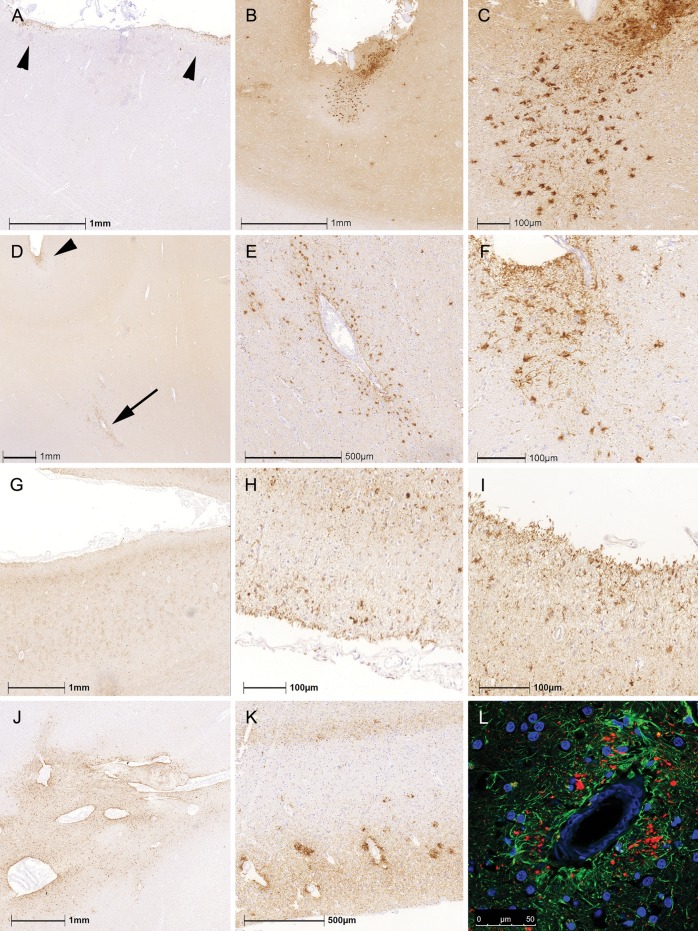
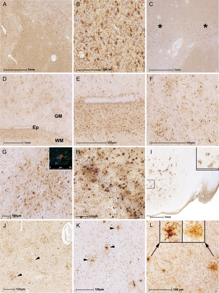
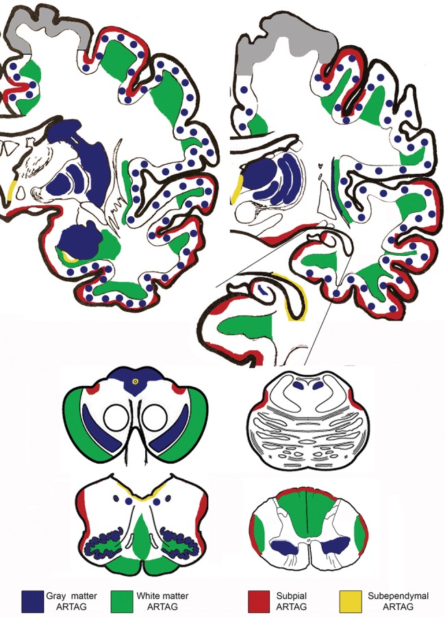
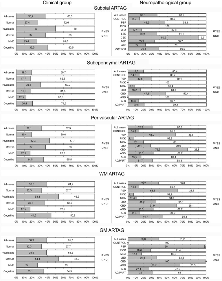
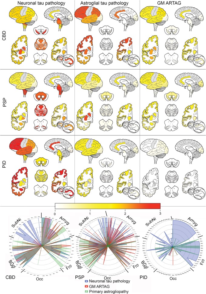
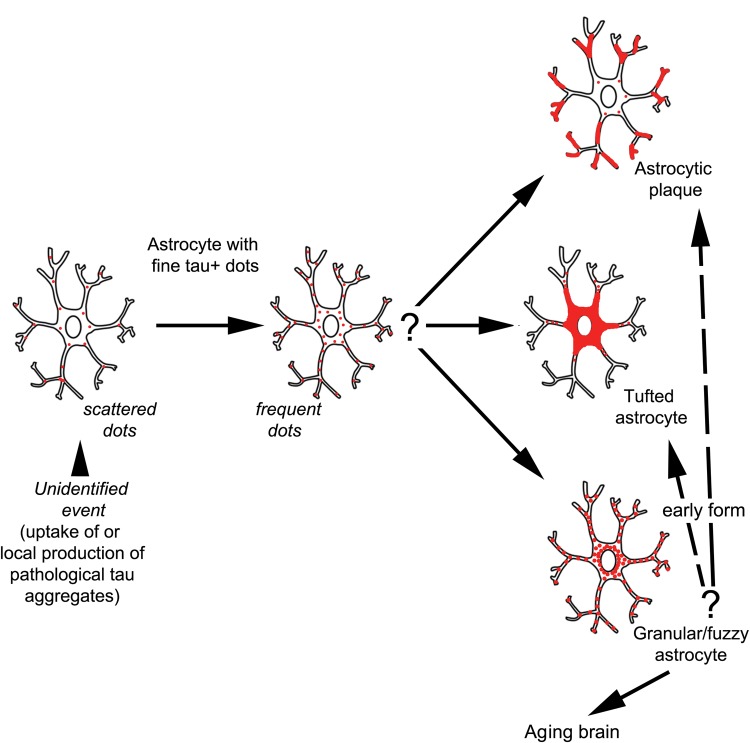
The term "aging-related tau astrogliopathy" (ARTAG) describes pathological accumulation of abnormally phosphorylated tau protein in astrocytes. We evaluated the correlates of ARTAG types (i.e., subpial, subependymal, white and gray matter, and perivascular) in different neuroanatomical regions. Clinical, neuropathological, and genetic (eg, APOE ε4 allele, MAPT H1/H2 haplotype) data from 628 postmortem brains from subjects were investigated; most of the patients had been longitudinally followed at the University of Pennsylvania. We found that (i) the amygdala is a hotspot for all ARTAG types; (ii) age at death, male sex, and presence of primary frontotemporal lobar degeneration (FTLD) tauopathy are significantly associated with ARTAG; (iii) age at death, greater degree of brain atrophy, ventricular enlargement, and Alzheimer disease (AD)-related variables are associated with subpial, white matter, and perivascular ARTAG types; (iv) AD-related variables are associated particularly with lobar white matter ARTAG; and (v) gray matter ARTAG in primary FTLD-tauopathies appears in areas without neuronal tau pathology. We provide a reference map of ARTAG types and propose at least 5 constellations of ARTAG. Furthermore, we propose a conceptual link between primary FTLD-tauopathy and ARTAG-related astrocytic tau pathologies. Our observations serve as a basis for etiological stratification and definition of progression patterns of ARTAG.

Keywords: ARTAG; Aging-related tau astrogliopathy; Alzheimer disease; Chronic traumatic encephalopathy; Dementia; Tau; Tauopathy.


Figures

FIGURE 1
FIGURE 1
Patterns of subpial and perivascular ARTAG. (AC) Distinct foci of subpial thorn-shaped astrocytes (TSA) in the anterior cingulate cortex in a patient with PSP. (DF) Perivascular ARTAG (D; indicated by an arrow and enlarged in E) was rarely associated with lobar subpial ARTAG (D; indicated by an arrowhead and enlarged in F). (GI) In CBD, we observed subpial tau-positive astrocytic processes with less prominent cytoplasmic tau immunoreactivity in the parietal lobe (G; enlarged in H and I). Perivascular ARTAG in the basal forebrain as exemplified in a PSP case (J). Accumulations of tau immunoreactivity around vessels showing amyloid angiopathy in AD (K) were related to dystrophic neuritic components (L, green represents GFAP, red phospho-tau, immunoreactivity).
FIGURE 2
FIGURE 2
Patterns of white and gray matter ARTAG. (AC) White matter ARTAG was characterized by thorn-shaped astrocytes in many brain regions (A, B) as well as in focal accentuations (C; indicated by asterisks). (DF) White matter (WM) ARTAG was associated frequently with perivascular, subependymal (D; indicated by Ep and enlarged in E) ARTAG and also with gray matter (GM) ARTAG (D; enlarged in F). (GL) Gray matter ARTAG was represented mostly by granular/fuzzy astrocytes (GFA) or TSA. In the brainstem the cytoplasm was plump (G; inset shows double immunolabeling for GFAP indicated by green and phospho-tau indicated by red). Accumulation of TSA in the dentate gyrus and CA4 in a case with clinically probable AD (H). Clusters of GFA in the amygdala in a case with schizophrenia (I; inset shows enlarged area). GFA-like astrocytic tau immunoreactivities were seen in CBD (J; parietal cortex), PSP (K; here frontal cortex), and in PiD (L; here occipital cortex: Left enlarged inset indicates a typical ramified astrocyte and the right, an enlarged image a GFA-type morphology).
FIGURE 3
FIGURE 3
Distribution patterns of ARTAG in this study. Note that perivascular is not indicated here because it was seen together with white matter ARTAG. Gray color in the cortex indicates areas that were not evaluated.
FIGURE 4
FIGURE 4
Percent of cases with (gray shading) or without (no shading) ARTAG types in clinical and neuropathological diagnostic groups. MND, motor neuron disease; MovDis, movement disorder (e.g. parkinsonism, corticobasal syndrome, PSP syndrome).
FIGURE 5
FIGURE 5
Heat maps and anatomical chart representation of tau pathologies in primary FTLD-tauopathies. Gray color indicates areas that were not systematically evaluated. In the radar chart, 5 anatomical regions are highlighted in all examined cases of PSP (n = 49), CBD (n = 21), and PiD (n = 14). Note that in the occipital cortex astroglial tau pathology predominates; furthermore, the proportions of astroglial (gray matter ARTAG or primary FTLD-tauopathy-related astrogliopathy, such as tufted astrocytes, astrocytic plaques and ramified astrocytes) and neuronal tau pathology differ considerably among PSP, CBD, and PiD cases.
FIGURE 6
FIGURE 6
Peculiar patterns of ARTAG. (AC) Gray and white matter ARTAG with thorny astrocytes in the anterior horn of the spinal cord (A; arrow indicates anterior horn enlarged in B; arrowhead indicates the anterior pyramidal tract enlarged in C). (DF) Widespread white and gray matter ARTAG in the medulla oblongata in an 81-year-old man with atypical parkinsonism (D; arrow indicates medial lemniscus on the left and hilum of the inferior olive on the right, enlarged in E; arrowhead indicates hypoglossal nucleus enlarged in F). (GL) Combined presence of globular oligodendroglial inclusions and thorn-shaped astrocytes in the peri-amygdaloid white matter (G; globular inclusions indicated by an arrowhead and enlarged in H and ARTAG indicated by an arrow and enlarged in I) and in the basal forebrain (J; perivascular ARTAG indicated by an arrowhead and enlarged in K and oligodendroglial globular inclusions indicated by an arrow and enlarged in L).
FIGURE 7
FIGURE 7
Summary of the concept of astroglial tau pathology in the gray matter. Scattered and frequent dots in astrocytic processes can be seen in various primary FTLD-tauopathies and in the aging brain. Granular/fuzzy astrocytes (GFA) can also be seen in the aging brain and in primary FTLD-tauopathies, while in CBD there are astrocytic plaques, and in PSP there are tufted astrocytes. This concept suggests that the fine dot-like astrocytic tau-immunoreactivity can progress to any of these morphologies and in some circumstances together with the GFA might represent the earliest steps of pathological tau accumulation in astrocytes.

References

    1. Kovacs GG, Ferrer I, Grinberg LT, et al. Aging-related tau astrogliopathy (ARTAG): harmonized evaluation strategy. Acta Neuropathol 2016;131:87–102 - PMC - PubMed
    1. Kovacs GG, Milenkovic I, Wohrer A, et al. Non-Alzheimer neurodegenerative pathologies and their combinations are more frequent than commonly believed in the elderly brain: a community-based autopsy series. Acta Neuropathol 2013;126:365–84 - PubMed
    1. Kovacs GG, Molnar K, Laszlo L, et al. A peculiar constellation of tau pathology defines a subset of dementia in the elderly. Acta Neuropathol 2011;122:205–22 - PubMed
    1. Munoz DG, Woulfe J, Kertesz A.. Argyrophilic thorny astrocyte clusters in association with Alzheimer's disease pathology in possible primary progressive aphasia. Acta Neuropathol 2007;114:347–57 - PubMed
    1. Ikeda K, Akiyama H, Kondo H, et al. Thorn-shaped astrocytes: possibly secondarily induced tau-positive glial fibrillary tangles. Acta Neuropathol 1995;90:620–5 - PubMed

MeSH terms