NLRP2 is a suppressor of NF-ƙB signaling and HLA-C expression in human trophoblasts†,‡
- PMID: 28340094
- PMCID: PMC5803763
- DOI: 10.1093/biolre/iox009
NLRP2 is a suppressor of NF-ƙB signaling and HLA-C expression in human trophoblasts†,‡
Abstract
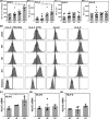
During pregnancy, fetal extravillous trophoblasts (EVT) play a key role in the regulation of maternal T cell and NK cell responses. EVT display a unique combination of human leukocyte antigens (HLA); EVT do not express HLA-A and HLA-B, but do express HLA-C, HLA-E, and HLA-G. The mechanisms establishing this unique HLA expression pattern have not been fully elucidated. The major histocompatibility complex (MHC) class I and class II transcriptional activators NLRC5 and CIITA are expressed neither by EVT nor by the EVT model cell line JEG3, which has an MHC expression pattern identical to that of EVT. Therefore, other MHC regulators must be present to control HLA-C, HLA-E, and HLA-G expression in these cells. CIITA and NLRC5 are both members of the nucleotide-binding domain, leucine-rich repeat (NLR) family of proteins. Another member of this family, NLRP2, is highly expressed by EVT and JEG3, but not in maternal decidual stromal cells. In this study, transcription activator-like effector nuclease technology was used to delete NLRP2 in JEG3. Furthermore, lentiviral delivery of shRNA was used to knockdown NLRP2 in JEG3 and primary EVT. Upon NLRP2 deletion, Tumor Necrosis Factor-α (TNFα)-induced phosphorylation of NF-KB p65 increased in JEG3 and EVT, and more surprisingly a significant increase in constitutive HLA-C expression was observed in JEG3. These data suggest a broader role for NLR family members in the regulation of MHC expression during inflammation, thus forming a bridge between innate and adaptive immune responses. As suppressor of proinflammatory responses, NLRP2 may contribute to preventing unwanted antifetal responses.
Keywords: HLA-G; MHC; NOD-like receptor; inflammasome; pregnancy.
© The Authors 2017. Published by Oxford University Press on behalf of Society for the Study of Reproduction. All rights reserved. For permissions, please journals.permissions@oup.com.
Figures


Similar articles
-
The Dual Role of HLA-C in Tolerance and Immunity at the Maternal-Fetal Interface.Front Immunol. 2019 Dec 9;10:2730. doi: 10.3389/fimmu.2019.02730. eCollection 2019. Front Immunol. 2019. PMID: 31921098 Free PMC article. Review.
-
HLA-C expression in extravillous trophoblasts is determined by an ELF3-NLRP2/NLRP7 regulatory axis.Proc Natl Acad Sci U S A. 2024 Jul 30;121(31):e2404229121. doi: 10.1073/pnas.2404229121. Epub 2024 Jul 25. Proc Natl Acad Sci U S A. 2024. PMID: 39052836 Free PMC article.
-
ELF3 activated by a superenhancer and an autoregulatory feedback loop is required for high-level HLA-C expression on extravillous trophoblasts.Proc Natl Acad Sci U S A. 2021 Mar 2;118(9):e2025512118. doi: 10.1073/pnas.2025512118. Proc Natl Acad Sci U S A. 2021. PMID: 33622787 Free PMC article.
-
Cytotoxic potential of decidual NK cells and CD8+ T cells awakened by infections.J Reprod Immunol. 2017 Feb;119:85-90. doi: 10.1016/j.jri.2016.08.001. Epub 2016 Aug 2. J Reprod Immunol. 2017. PMID: 27523927 Free PMC article. Review.
-
The effect of heat shock protein 27 on extravillous trophoblast differentiation and on eukaryotic translation initiation factor 4E expression.Mol Hum Reprod. 2014 May;20(5):422-32. doi: 10.1093/molehr/gau002. Epub 2014 Jan 14. Mol Hum Reprod. 2014. PMID: 24431103
Cited by
-
The NLR gene family: from discovery to present day.Nat Rev Immunol. 2023 Oct;23(10):635-654. doi: 10.1038/s41577-023-00849-x. Epub 2023 Mar 27. Nat Rev Immunol. 2023. PMID: 36973360 Free PMC article. Review.
-
NLRP7 is increased in human idiopathic fetal growth restriction and plays a critical role in trophoblast differentiation.J Mol Med (Berl). 2019 Mar;97(3):355-367. doi: 10.1007/s00109-018-01737-x. Epub 2019 Jan 8. J Mol Med (Berl). 2019. PMID: 30617930
-
Inflammasomes-A Molecular Link for Altered Immunoregulation and Inflammation Mediated Vascular Dysfunction in Preeclampsia.Int J Mol Sci. 2020 Feb 19;21(4):1406. doi: 10.3390/ijms21041406. Int J Mol Sci. 2020. PMID: 32093005 Free PMC article. Review.
-
The Dual Role of HLA-C in Tolerance and Immunity at the Maternal-Fetal Interface.Front Immunol. 2019 Dec 9;10:2730. doi: 10.3389/fimmu.2019.02730. eCollection 2019. Front Immunol. 2019. PMID: 31921098 Free PMC article. Review.
-
Role of Microgliosis and NLRP3 Inflammasome in Parkinson's Disease Pathogenesis and Therapy.Cell Mol Neurobiol. 2022 Jul;42(5):1283-1300. doi: 10.1007/s10571-020-01027-6. Epub 2021 Jan 2. Cell Mol Neurobiol. 2022. PMID: 33387119 Free PMC article. Review.
References
-
- Moffett-King A. Natural killer cells and pregnancy. Nat Rev Immunol 2002; 2:656–663. - PubMed
-
- Kovats S, Main EK, Librach C, Stubblebine M, Fisher SJ, DeMars R. A class I antigen, HLA-G, expressed in human trophoblasts. Science 1990; 248:220–223. - PubMed
-
- Holling TM, Bergevoet MW, Wierda RJ, van Eggermond MC, van den Elsen PJ. Genetic and epigenetic control of the major histocompatibility complex class Ib gene HLA-G in trophoblast cell lines. Ann NY Acad Sci 2009; 1173:538–544. - PubMed
MeSH terms
Substances
LinkOut - more resources
Full Text Sources
Other Literature Sources
Research Materials

