. 2017 Mar 23;169(1):72-84.e13.
doi: 10.1016/j.cell.2017.03.007.

Human Epistatic Interaction Controls IL7R Splicing and Increases Multiple Sclerosis Risk

Affiliations

Human Epistatic Interaction Controls IL7R Splicing and Increases Multiple Sclerosis Risk

Gaddiel Galarza-Muñoz et al. Cell. .

Abstract

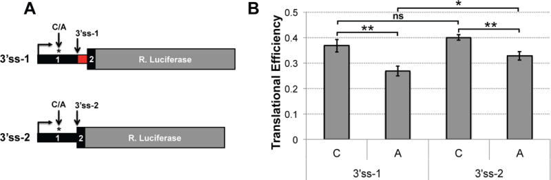
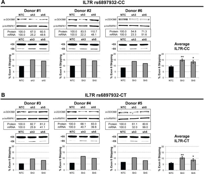
Multiple sclerosis (MS) is an autoimmune disorder where T cells attack neurons in the central nervous system (CNS) leading to demyelination and neurological deficits. A driver of increased MS risk is the soluble form of the interleukin-7 receptor alpha chain gene (sIL7R) produced by alternative splicing of IL7R exon 6. Here, we identified the RNA helicase DDX39B as a potent activator of this exon and consequently a repressor of sIL7R, and we found strong genetic association of DDX39B with MS risk. Indeed, we showed that a genetic variant in the 5' UTR of DDX39B reduces translation of DDX39B mRNAs and increases MS risk. Importantly, this DDX39B variant showed strong genetic and functional epistasis with allelic variants in IL7R exon 6. This study establishes the occurrence of biological epistasis in humans and provides mechanistic insight into the regulation of IL7R exon 6 splicing and its impact on MS risk.

Keywords: DDX39B; IL7R; alternative splicing; autoimmune disorders; epistasis; genetic association; multiple sclerosis.


Figures

Figure 1
Figure 1. DDX39B regulates alternative splicing of IL7R exon 6
A–D, Knockdown of DDX39B in HeLa cells using two independent DDX39B siRNAs (DDX_3 and DDX_4) and a non-silencing control siRNA (NSC). A, Western blot analysis illustrating depletion of DDX39B. BC, RT-PCR analysis of IL7R exon 6 splicing (+E6 = exon included; −E6 = exon skipped) in transcripts from a reporter minigene (B) or the endogenous gene (C). D, Quantification of sIL7R secretion by ELISA. E–F, Rescue experiments with HeLa cell lines stably expressing siRNA-resistant DDX39B trans-gene, either wild type (E, WT) or helicase mutant (F, D199A), under the control of the tetracycline operator. Top panels illustrate DDX39B western blot analysis, whereas lower panels show RT-PCR analysis of endogenous IL7R transcripts. In all panels the data is shown as mean ± s.d., and statistical significance was assessed using Student’s t-test (*** p0.0005; ** p0.005; * p0.05). See also Figures S2 and S3.
Figure 2
Figure 2. Variants within or adjacent to the DDX39B locus strongly associated with MS risk
Each diamond represents a variant analyzed, and the color of the diamonds indicates no association (gray), marginal association (yellow) or strong association (red) with MS risk. The variant rs2523506 is indicated with a larger diamond (see Figure 3). Black and blue dotted lines indicate thresholds for marginal (p ≤ 1.0 × 10²) and strong (p ≤ 5.0 × 10⁸) association, respectively. The location of the four genes present in this region of chromosome 6 is illustrated at the bottom. See also Figure S5 and Table S3.
Figure 3
Figure 3. The DDX39B 5′ UTR variant rs2523506 displays allele-specific DDX39B protein expression
A, Schematic representation of the DDX39B gene (black), spliced mRNA isoforms (red) and location of MS-associated variants rs2523506, rs2523512 and rs2516478 (asterisks). B, RT-qPCR quantification of DDX39B mRNA levels in human PBMCs (left) and African (YRI/ESN, middle) and European (IBS, right) LCLs stratified by rs2523506 genotype. Each symbol represents cells from one individual, and red lines indicate median and interquartile range for each group. Samples sizes were: PBMC, CC=32, AC=31, AA=23; YRI/ESN, CC=12, AC=11, AA=2; and IBS, CC=12, AC=12, AA=3. C, Western blot analysis of DDX39B protein abundance in African (YRI/ESN, left) and European (IBS, right) LCLs stratified by rs2523506 genotype. Panels in C were assembled with different portions of the same gel. Statistical significance of all measurements was assessed using Student’s t-test (two-sided; *** p0.0005; ** p0.005; * p0.05). See also Figures S7, S8 and S9.
Figure 4
Figure 4. The risk allele of rs2523506 reduces translational efficiency mediated by DDX39B 5′ UTRs
A, Schematic representation of the different DDX39B 5′ UTR luciferase reporters, which differ by alternative 3′ss in exon 2 and the single nucleotide change at rs2523506 (C/A). B, Measurements of translational efficiency in transfected HeLa cells. RNA levels and luciferase activity were measured by RT-qPCR and dual Luciferase assays, respectively. Translational efficiency was determined by dividing luciferase activity by RNA levels. Statistical significance was assessed using Student’s t-test (two-sided; ** p0.005; * p0.05).
Figure 5
Figure 5. Functional interaction between DDX39B and IL7R exon 6 variants
The interaction between DDX39B and IL7R was functionally tested in DDX39B-depleted HeLa cells using IL7R splicing reporters carrying either the risk C allele (IL7R-C; left) or the protective T allele (IL7R-T; right) of rs6897932. RT-PCR analysis of exon 6 splicing (mean ± s.d.) reveals higher exon 6 skipping when the levels of DDX39B are reduced in the context of the risk C allele than of the protective T allele of rs6897932 (Student’s t-test, two-sided; ** p0.005).
Figure 6
Figure 6. DDX39B regulates IL7R exon 6 splicing in primary CD4+ T cells
Knockdown of DDX39B in primary CD4+ T cells from six donors via lentiviral transduction with two independent shRNA against DDX39B (sh3 and sh5) and a non-targeting control shRNA (NTC). Donors are grouped by IL7R genotype: IL7R-CC (A) and IL7R-CT (B), and each panel illustrates DDX39B western blot analysis (top) and RT-PCR analysis of IL7R exon 6 splicing (bottom) for each donor individually. In each panel, the plots on the right show the average of % exon 6 skipping for each IL7R genotype (mean ± s.d., Student’s t-test: ** p0.005; * p0.05).

References

    1. Allcock RJ, Williams JH, Price P. The central MHC gene, BAT1, may encode a protein that down-regulates cytokine production. Genes Cells. 2001;6:487–494. - PubMed
    1. Alves NL, van Leeuwen EM, Derks IA, van Lier RA. Differential regulation of human IL-7 receptor alpha expression by IL-7 and TCR signaling. Journal of immunology. 2008;180:5201–5210. - PubMed
    1. Anderson CA, Boucher G, Lees CW, Franke A, D’Amato M, Taylor KD, Lee JC, Goyette P, Imielinski M, Latiano A, et al. Meta-analysis identifies 29 additional ulcerative colitis risk loci, increasing the number of confirmed associations to 47. Nature genetics. 2011;43:246–252. - PMC - PubMed
    1. Badot V, Durez P, Van den Eynde BJ, Nzeusseu-Toukap A, Houssiau FA, Lauwerys BR. Rheumatoid arthritis synovial fibroblasts produce a soluble form of the interleukin-7 receptor in response to pro-inflammatory cytokines. J Cell Mol Med. 2011;15:2335–2342. - PMC - PubMed
    1. Barrett JC, Clayton DG, Concannon P, Akolkar B, Cooper JD, Erlich HA, Julier C, Morahan G, Nerup J, Nierras C, et al. Genome-wide association study and meta-analysis find that over 40 loci affect risk of type 1 diabetes. Nature genetics. 2009;41:703–707. - PMC - PubMed

Publication types