. 2017 Mar 24;10(1):18.
doi: 10.1186/s12920-017-0249-2.

Gene and protein analysis reveals that p53 pathway is functionally inactivated in cytogenetically normal Acute Myeloid Leukemia and Acute Promyelocytic Leukemia

Affiliations

Gene and protein analysis reveals that p53 pathway is functionally inactivated in cytogenetically normal Acute Myeloid Leukemia and Acute Promyelocytic Leukemia

Julia Abramowitz et al. BMC Med Genomics. .

Abstract

Background: Mechanisms that inactivate the p53 pathway in Acute Myeloid Leukemia (AML), other than rare mutations, are still not well understood.

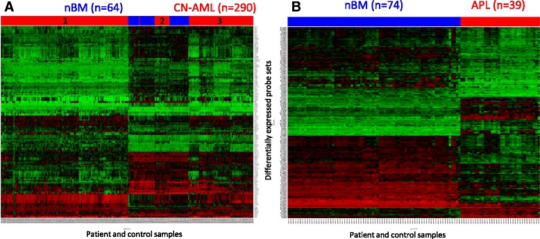
Methods: We performed a bioinformatics study of the p53 pathway function at the gene expression level on our collection of 1153 p53-pathway related genes. Publically available Affymetrix data of 607 de-novo AML patients at diagnosis were analyzed according to the patients cytogenetic, FAB and molecular mutations subtypes. We further investigated the functional status of the p53 pathway in cytogenetically normal AML (CN-AML) and Acute Promyelocytic Leukemia (APL) patients using bioinformatics, Real-Time PCR and immunohistochemistry.

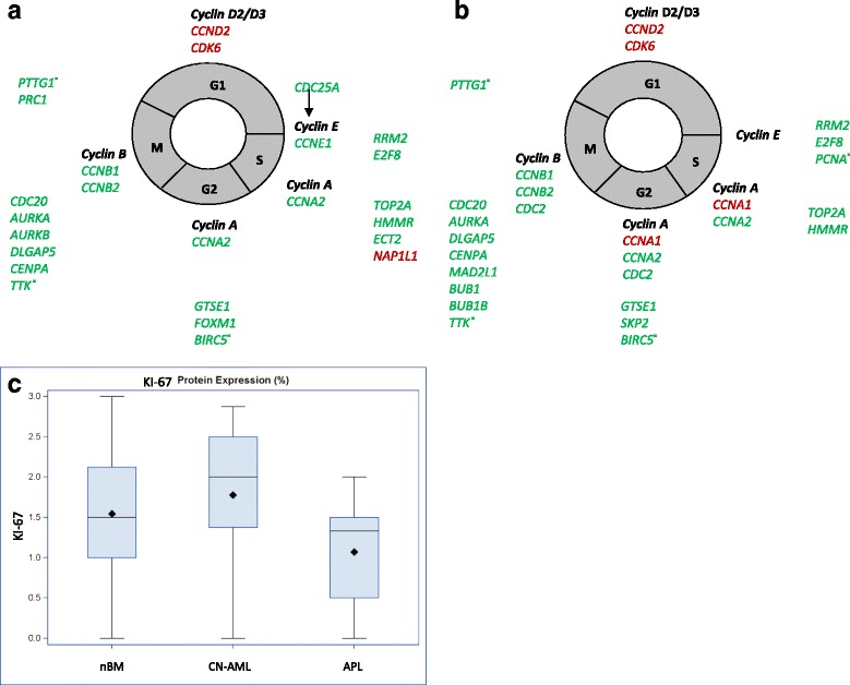
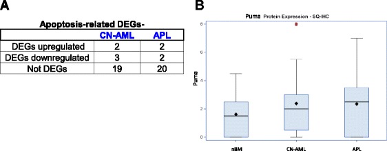
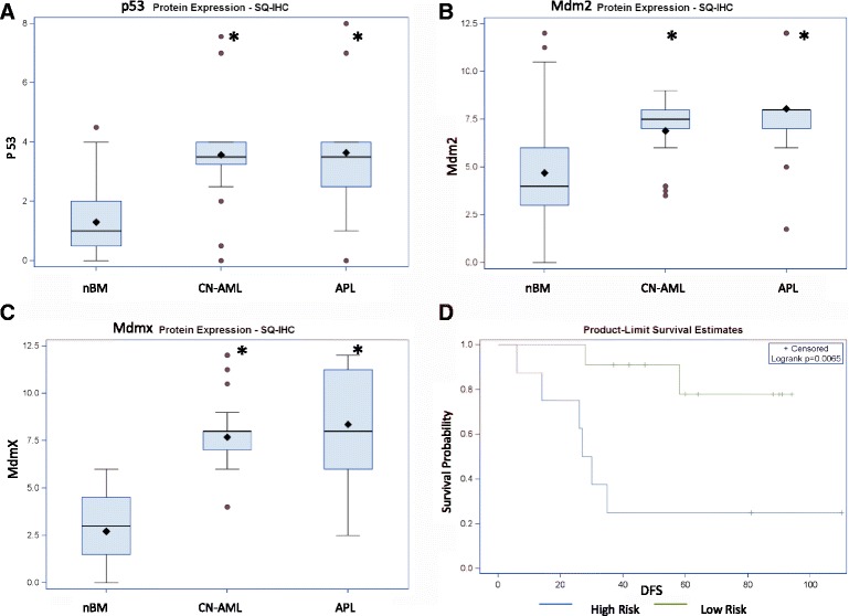
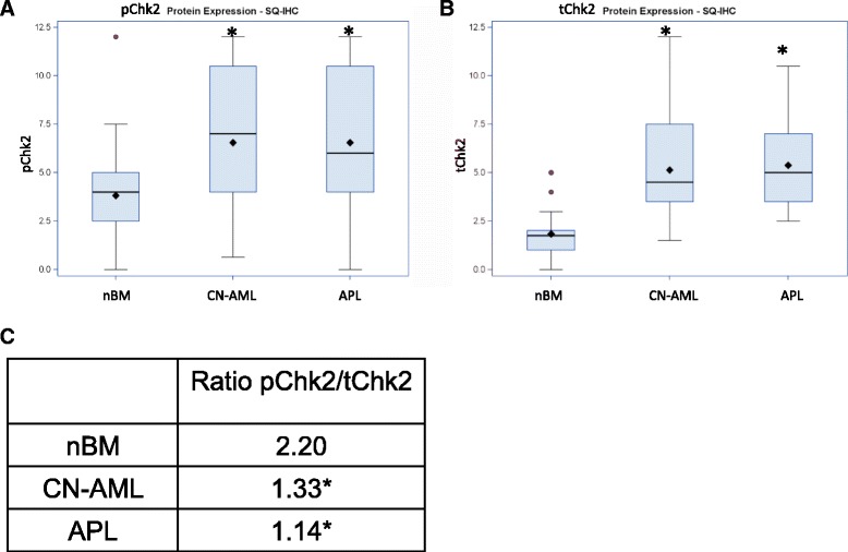
Results: We revealed significant and differential alterations of p53 pathway-related gene expression in most of the AML subtypes. We found that p53 pathway-related gene expression was not correlated with the accepted grouping of AML subtypes such as by cytogenetically-based prognosis, morphological stage or by the type of molecular mutation. Our bioinformatic analysis revealed that p53 is not functional in CN-AML and APL blasts at inducing its most important functional outcomes: cell cycle arrest, apoptosis, DNA repair and oxidative stress defense. We revealed transcriptional downregulation of important p53 acetyltransferases in both CN-AML and APL, accompanied by increased Mdmx protein expression and inadequate Chk2 protein activation.

Conclusions: Our bioinformatic analysis demonstrated that p53 pathway is differentially inactivated in different AML subtypes. Focused gene and protein analysis of p53 pathway in CN-AML and APL patients imply that functional inactivation of p53 protein can be attributed to its impaired acetylation. Our analysis indicates the need in further accurate evaluation of p53 pathway functioning and regulation in distinct subtypes of AML.


Figures

Fig. 1
Fig. 1
Heat maps of differentially expressed p53-related probe sets in CN-AML and APL. a CN-AML, b APL. Red -upregulation of gene expression, green-downregulation of gene expression, black-no change in gene expression. Numbers of enrolled patients (red) and controls (blue) are indicated on top and color coded. Subgroups of CN-AML patients are designated as 1, 2 and 3
Fig. 2
Fig. 2
Cell cycle in CN-AML and APL. a, b DEGs that are typically repressed by p53 and are associated with the cell cycle in CN-AML (a) and APL (b). Canonical cyclins in each phase of the cell cycle are in bold black. DEGs implicated in the different phases of the cell cycle are placed near the appropriate phase. Upregulated DEGs are in red and downregulated DEGs are in green. Most of the DEGs arrest proliferation, while only CCND2, CDK6 and NAP1L1 genes support proliferation. Most of the genes originate from 2 gene expression signatures [71, 72], Additional 13 genes are marked withˣ. CCNE1 (a) is the only DEG not repressed by p53. c Percent of KI-67 positive cells. Immunohistochemistry was performed on bone marrow samples of 25 CN-AML and 23 APL patients at diagnosis and 35 nBM samples. The boundaries of the blue box indicate the 25th percentile (bottom boundary) and the 75th percentile (top boundary) of KI-67 level, median is displayed by the thick line in the box, mean by rhombus sign
Fig. 3
Fig. 3
DEGs associated with apoptosis. a Apoptosis-related DEGs in CN-AML and APL among canonical p53-dependent pro-apoptotic genes [75]. We extended some gene families (e.g. PIGs) and the resultant gene collection includes 24 genes. The table summarizes the numbers of genes that were found to be upregulated DEGs, downregulated DEGs or not identified as DEGs in our study. b Puma protein levels by IHC in nBM, and in CN-AML and APL patients’ BM. Symbols of box plots are as in Fig. 2; outlying value is marked by red circle; tails of the distribution depicted only in one direction indicate that the values are skewed towards that side of the average
Fig. 4
Fig. 4
p53, Mdm2 and Mdmx protein levels in nBM, CN-AML and APL. a P53 protein, b Mdm2 protein, c Mdmx protein; protein levels are according to IHC ( SQ score); *-p-value < 0.05 (compared to nBM); Box plots symbols are as in Fig. 2 and 3. d Kaplan-Meier survival curve of APL patients according to Mdmx protein levels. Mdmx high risk group (blue) n = 8, low risk (green) n = 11. DFS- disease free survival
Fig. 5
Fig. 5
pChk2 and tChk2 protein levels in nBM, CN-AML and APL. a pChk2 protein, b tChk2 protein levels by SQ score. Box plots symbols are as in Fig. 2 and 3. c ratio of pChk2 to tChk2 by the median SQ score. *-p-value < 0.05 (compared to normal bone marrow)

Similar articles

Cited by

References

    1. Acute Myeloid Leukemia. Leukemia and Lymphoma Society. 2015. www.sllcanada.org/sites/default/files/file_assets/PS32_AML_Booklet_FINAL....
    1. Foran JM. New prognostic markers in acute myeloid leukemia: perspective from the clinic. Hematology Am Soc Hematol Educ Program. 2010;2010:47–55. - PubMed
    1. Renneville A, Roumier C, Biggio V, Nibourel O, Boissel N, Fenaux P, et al. Cooperating gene mutations in acute myeloid leukemia: a review of the literature. Leukemia. 2008;22(5):915–31. doi: 10.1038/leu.2008.19. - DOI - PubMed
    1. Theilgaard-Monch K, Boultwood J, Ferrari S, Giannopoulos K, Hernandez-Rivas JM, Kohlmann A, et al. Gene expression profiling in MDS and AML: potential and future avenues. Leukemia. 2011;25(6):909–20. doi: 10.1038/leu.2011.48. - DOI - PubMed
    1. Bullinger L, Dohner K, Bair E, Frohling S, Schlenk RF, Tibshirani R, et al. Use of gene-expression profiling to identify prognostic subclasses in adult acute myeloid leukemia. N Engl J Med. 2004;350(16):1605–16. doi: 10.1056/NEJMoa031046. - DOI - PubMed

Publication types

MeSH terms

Substances