. 2017 Mar 24;14(1):62.
doi: 10.1186/s12974-017-0836-3.

Lymphocytes have a role in protection, but not in pathogenesis, during La Crosse Virus infection in mice

Affiliations

Lymphocytes have a role in protection, but not in pathogenesis, during La Crosse Virus infection in mice

Clayton W Winkler et al. J Neuroinflammation. .

Abstract

Background: La Crosse Virus (LACV) is a primary cause of pediatric viral encephalitis in the USA and can result in severe clinical outcomes. Almost all cases of LACV encephalitis occur in children 16 years or younger, indicating an age-related susceptibility. This susceptibility is recapitulated in a mouse model where weanling (3 weeks old or younger) mice are susceptible to LACV-induced disease, and adults (greater than 6 weeks) are resistant. Disease in mice and humans is associated with infiltrating leukocytes to the CNS. However, what cell types are infiltrating into the brain during virus infection and how these cells influence pathogenesis remain unknown.

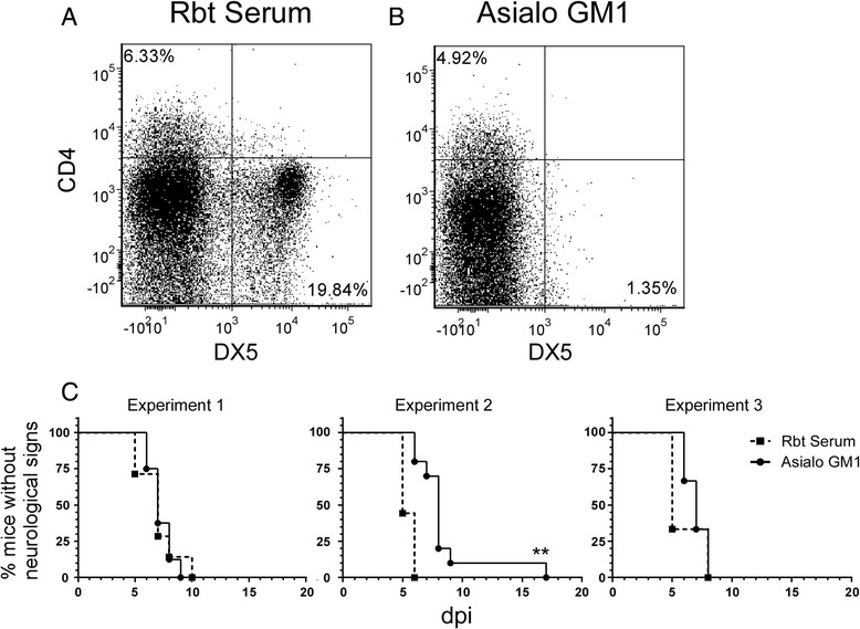
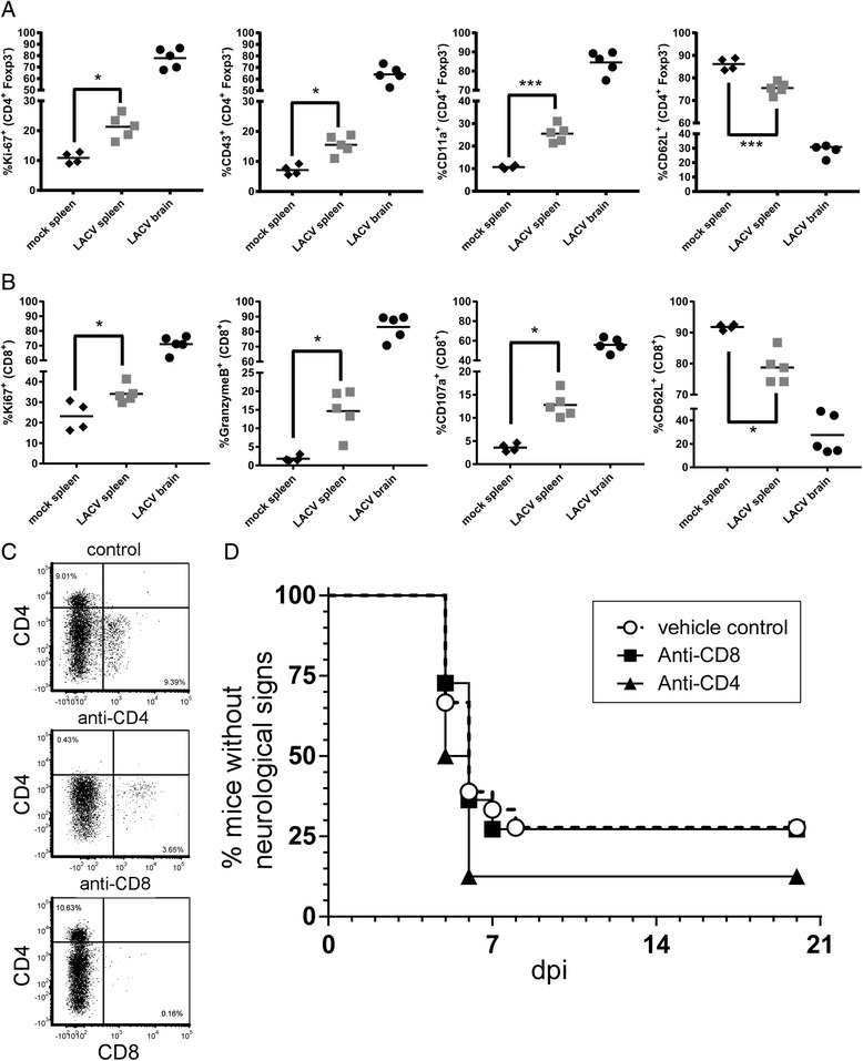
Methods: In the current study, we analyzed lymphocytes recruited to the CNS during LACV-infection in clinical mice, using flow cytometry. We analyzed the contribution of these lymphocytes to LACV pathogenesis in weanling mice using knockout mice or antibody depletion. Additionally, we studied at the potential role of these lymphocytes in preventing LACV neurological disease in resistant adult mice.

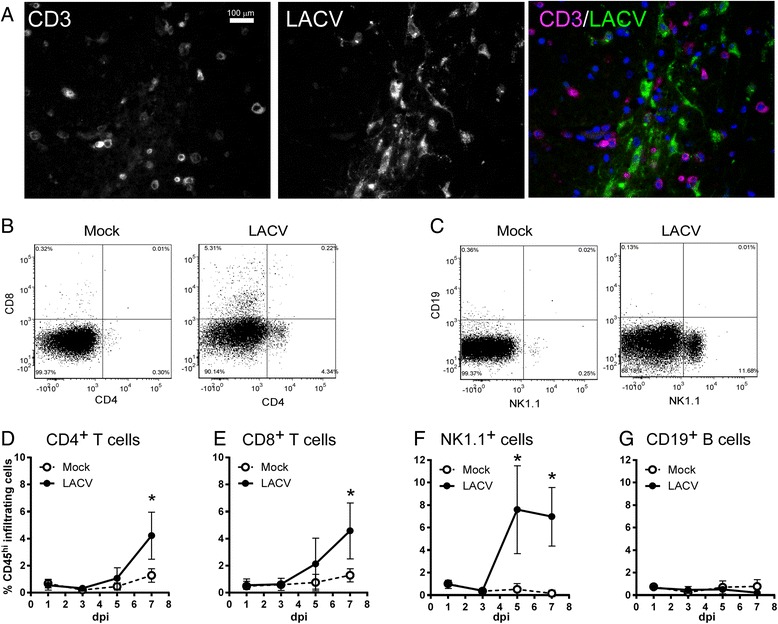
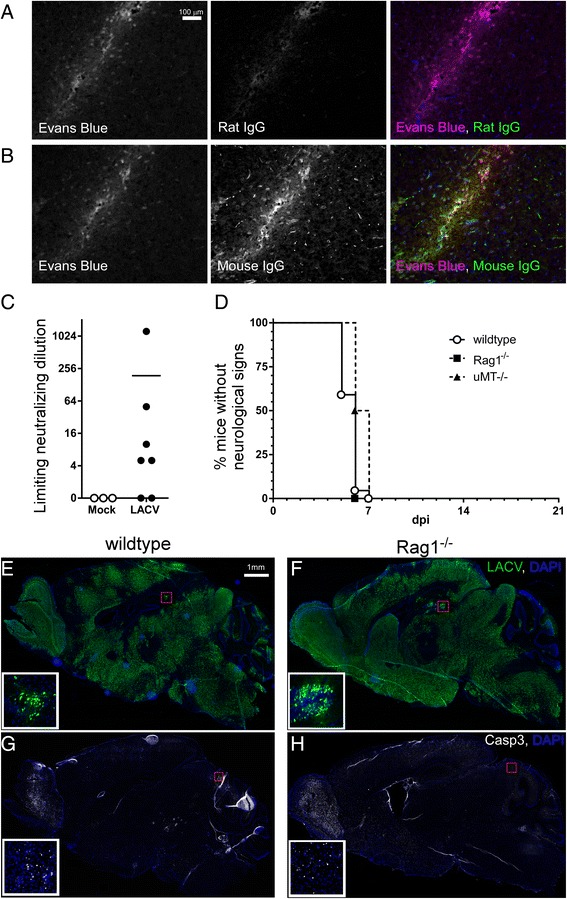
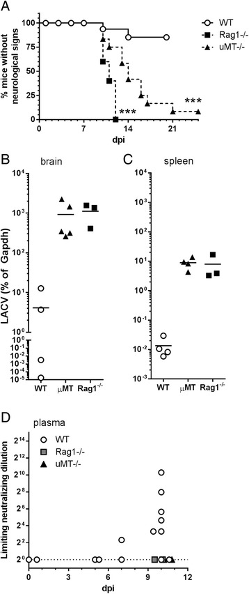
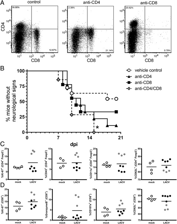
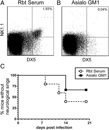
Results: In susceptible weanling mice, disease was associated with infiltrating lymphocytes in the CNS, including NK cells, CD4 T cells, and CD8 T cells. Surprisingly, depletion of these cells did not impact neurological disease, suggesting these cells do not contribute to virus-mediated damage. In contrast, in disease-resistant adult animals, depletion of both CD4 T cells and CD8 T cells or depletion of B cells increased neurological disease, with higher levels of virus in the brain.

Conclusions: Our current results indicate that lymphocytes do not influence neurological disease in young mice, but they have a critical role protecting adult animals from LACV pathogenesis. Although LACV is an acute virus infection, these studies indicate that the innate immune response in adults is not sufficient for protection and that components of the adaptive immune response are necessary to prevent virus from invading the CNS.

Keywords: Brain; Central nervous system; Encephalitis; La Crosse Virus; Lymphocytes.


Figures

Fig. 1
Fig. 1
Lymphocyte infiltration into the CNS following LACV infection of weanling mice. a Brain tissue sections from an LACV-infected mouse at a clinical time point (7 dpi) was stained for anti-CD3 (first panel, third panel: magenta) and LACV (second panel, third panel: green). bg Analysis of infiltrating cells by flow cytometry. Brain tissue was removed from mock or LACV-infected mice at 1, 3, 5, or 7 dpi and immune cells were isolated, antibody labeled, and analyzed by flow cytometry as indicated in the “Methods”. Infiltrating cells were gated by CD45 high expression (data not shown) and then for b CD4 and CD8 expression as well as c CD19 and NK1.1 expression. b, c are representative data from mice at 7 dpi. dg Time course analysis of percent (%) infiltrating cell populations including d CD4+ cells, e CD8+ cells, f NK1.1+ cells, and g CD19+ B cells relative to the total number of live, CD45hi cells. Data are the mean ± SD for three to nine samples per time point for LACV-infected mice and one to three mice per time point for mock-infected controls. *P value <0.05 as determined by two-way ANOVA with Sidak’s multiple comparison test
Fig. 2
Fig. 2
NK cell depletion slightly delays onset of LACV-induced neurological disease in weanling mice. ac LACV-infected weanling mice were treated with a rabbit serum or b anti-Asialo GM1 to deplete NK cells as described in the “Methods.” Flow cytometry analysis at 7 dpi was used to confirm that treatment with b anti-Asialo GM1, but not a rabbit serum resulted in depletion of NK cells as shown by CD49b (DX5) expression. Experiment 1 included 4–5 mice per group, experiment 2 had 9–10 mice per group, and experiment 3 had 3 mice per group. Statistical analysis was completed using the Mantel-Cox log-rank test
Fig. 3
Fig. 3
CNS-infiltrating T cells are proliferating and activated during LACV infection but do not significantly alter LACV pathogenesis in weanling mice. Splenocytes and CNS-infiltrating a CD4+, Foxp3 T helper, and b CD8+ CD3+ cytotoxic T cells were analyzed for expression of proliferation and activation markers by flow cytometry at the clinical time point (6–7 dpi). Data are presented as percent (%) of either CD4+ or CD8+ T cells positive for the proliferation marker Ki-67, the activation markers CD43, CD11a, GranzymeB, or CD107a or the naïve T cell marker CD62L. c LACV-infected weanling mice were treated with RPMI/10% FBS (control), anti-CD4 or anti-CD8-depleting monoclonal antibodies as described in the “Methods”. Depletions were confirmed by flow cytometry analysis of brain tissue at the clinical time point (5–7 dpi). Data are a summary plot of two independent experiments with 4–12 mice per group. d 18 vehicle control-, 11 anti-CD8-, and 8 anti-CD4-treated mice were followed for the development of clinical signs of neurological disease. Statistical analysis was completed using the Mantel-Cox log-rank test
Fig. 4
Fig. 4
Immunoglobulin can be detected in the brain of LACV-infected weanling mice but does not influence the development of neurological disease or neuropathology. a, b Detection of antibody in the brain by immunohistochemistry of LACV-infected weanling mice at the clinical time point (6 dpi) that were treated i.p. with Evans Blue dye 1 h prior to tissue removal. Brain tissue sections were analyzed for Evans Blue to detect vascular leakage (magenta) and either a rat Ig as a negative control or b mouse IgG (green) was used to detect leakage of antibodies into the brain. c Analysis of plasma from LACV-infected weanling mice at 4–5 dpi for NAb. Data are plotted as the limiting dilution for the inhibition of virus replication on a log2 scale. Each symbol represents an individual animal. d Weanling wildtype (n = 22), μMT -/- (n = 6), and Rag1 -/- (n = 10) mice develop neurological disease with similar rate and frequency. All mice were 3 weeks of age when infected i.p. with 103 PFU of LACV and followed for clinical disease. Statistical analysis was completed using the Mantel-Cox log-rank test with no significant difference detected. Representative, adjacent sections of brain tissue from e, g LACV-infected weanling wildtype and f, h Rag1 -/- mice with neurological disease (6–7 dpi) were stained for LACV (green) and active-Caspase 3 (white). Higher magnification insets in e and f show infected cells (LACV: green, DAPI: blue) with neuronal morphology within the hippocampus (red boxes). Insets in g and h demonstrate cells undergoing apoptosis (active-Caspase 3, white) within similar regions of the midbrain (red boxes) in both genotypes
Fig. 5
Fig. 5
Adult Rag1 -/- and μMT -/- mice have increased susceptibility to LACV infection. ad Adult (6–8 weeks old) wildtype, Rag1 -/-, and μMT -/- mice were infected with 103 PFU of LACV and examined for a development of neurological disease, b, c virus RNA levels in the b brain and c spleen as well as the production of d neutralizing antibodies. a Data are presented as survival curve analysis of wildtype (n = 37), Rag1 -/- (n = 5), and μMT -/- (n = 12) mice. Statistical analysis was completed using the Mantel-Cox log-rank test. *** P <0.01. b, c RNA isolated from wildtype, Rag1 -/-, and μMT -/- mice at 10–12 dpi was assayed for LACV RNA by real-time PCR. Data were analyzed as described in “Methods” and are presented as a ratio of expression relative to housekeeping gene for each sample. Each symbol represents an individual mouse. d Neutralizing antibodies were detected at 5–10 dpi in wildtype mice. Data are presented as the dilution of plasma resulting in 50% inhibition of virus infection on a log2 scale. No detectable neutralizing antibodies were observed in Rag1 -/- or μMT -/- mice
Fig. 6
Fig. 6
CD4 and CD8 T cells contribute to protection against LACV-induced disease in adult mice. a, b LACV-infected adult mice were treated with RPMI/10% FBS as a vehicle control, anti-CD4, or anti-CD8 as described in the “Methods”. a CD4 and CD8 depletion were confirmed by flow cytometry analysis of splenocytes. b Neurological disease development from vehicle-treated controls (n = 11), anti-CD4-treated (n = 9), anti-CD8-treated (n = 9), and anti-CD4/anti-CD8-treated (n = 7) mice. Statistical analysis was completed using the Mantel-Cox log-rank test. Although individual anti-CD4 or anti-CD8-treated mice had slightly increased incidence of neurological disease, only depletion of both subsets resulted in a significant (* P < 0.05) increase in neurological disease. c CD4+, Foxp3 T helper, and d CD8+ CD3+ cytotoxic T cells from the spleens of adult mice at 7 dpi were analyzed for expression of proliferation and activation markers by flow cytometry. Data are presented as percent (%) of either CD4+ or CD8+ T cells positive for the proliferation marker Ki-67, the activation markers CD43, CD11a, GranzymeB, or CD107a or the naïve T cell marker CD62L. Gray circles indicate a group of three mice that showed consistent activation of CD4 and CD8 T cell responses, while black circles indicate mice that did not have increased responses
Fig. 7
Fig. 7
NK cell depletion does not affect susceptibility in adult mice. ac LACV-infected adult mice were treated with a rabbit serum or b anti-Asialo GM1 to deplete NK cells as described in the “Methods”. Flow cytometry analysis was used to confirm that treatment with b anti-Asialo GM1, but not a rabbit serum resulted in depletion of NK cells. c NK cell depletion did not significantly affect development of neurological disease. Data are plotted as a survival curve analysis of five rabbit serum-treated and six anti-Asialo GM1-treated mice. Statistical analysis was completed using the Mantel-Cox log-rank test. No statistical difference was observed

Similar articles

Cited by

References

    1. Thompson WH, Kalfayan B, Anslow RO. Isolation of California encephalitis group virus from a fatal human illness. Am J Epidemiol. 1965;81:245–253. doi: 10.1093/oxfordjournals.aje.a120512. - DOI - PubMed
    1. McJunkin JE, Nahata MC, De Los Reyes EC, Hunt WG, Caceres M, Khan RR, Chebib MG, Taravath S, Minnich LL, Carr R, et al. Safety and pharmacokinetics of ribavirin for the treatment of la crosse encephalitis. Pediatr Infect Dis J. 2011;30:860–865. doi: 10.1097/INF.0b013e31821c922c. - DOI - PubMed
    1. Westby KM, Fritzen C, Paulsen D, Poindexter S, Moncayo AC. La Crosse encephalitis virus infection in field-collected Aedes albopictus, Aedes japonicus, and Aedes triseriatus in Tennessee. J Am Mosq Control Assoc. 2015;31:233–241. doi: 10.2987/moco-31-03-233-241.1. - DOI - PubMed
    1. Schuh T, Schultz J, Moelling K, Pavlovic J. DNA-based vaccine against La Crosse virus: protective immune response mediated by neutralizing antibodies and CD4+ T cells. Hum Gene Ther. 1999;10:1649–1658. doi: 10.1089/10430349950017653. - DOI - PubMed
    1. Bennett RS, Gresko AK, Nelson JT, Murphy BR, Whitehead SS. A recombinant chimeric La Crosse virus expressing the surface glycoproteins of Jamestown Canyon virus is immunogenic and protective against challenge with either parental virus in mice or monkeys. J Virol. 2012;86:420–426. doi: 10.1128/JVI.02327-10. - DOI - PMC - PubMed

Publication types

LinkOut - more resources