. 2017 Mar 24;17(1):33.
doi: 10.1186/s12896-017-0353-6.

Stable high-level expression of factor VIII in Chinese hamster ovary cells in improved elongation factor-1 alpha-based system

Affiliations

Stable high-level expression of factor VIII in Chinese hamster ovary cells in improved elongation factor-1 alpha-based system

Nadezhda A Orlova et al. BMC Biotechnol. .

Abstract

Background: Recombinant factor VIII (FVIII), used for haemophilia A therapy, is one of the most challenging among the therapeutic proteins produced in heterologous expression systems. Deletion variant of FVIII, in which the entire domain B is replaced by a short linker peptide, was approved for medical use. Efficacy and safety of this FVIII deletion variant are similar to full-length FVIII preparations while the level of production in CHO cells is substantially higher. Typical levels of productivity for CHO cell lines producing deletion variant FVIII-BDD SQ, described elsewhere, are 0.5-2 IU/ml, corresponding to the concentration of FVIII of about 0.2 μg/ml. Using standard vectors based on the cytomegalovirus promoter (CMV) and the dihydrofolate reductase cDNA we have previously obtained the cell line secreting 0.5 IU/ml of FVIII-BDD, which roughly corresponds to the previously published data.

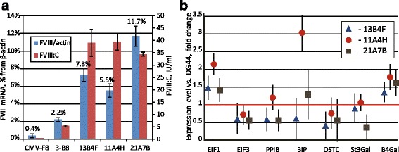
Results: An expression system based on CHO genomic sequences including CHO-EEF1A promoter and Epstein-Barr virus terminal repeat fragment allowed us to achieve 80-fold increase in the production level as compared with the conventional expression system based on the CMV promoter. Immediately after the primary selection FVIII -producing cells secreted 5-10 IU/ml of FVIII-BDD, and after multi-stage methotrexate-driven amplification a stable clonal line 11A4H was selected, secreting 39 IU/ml of FVIII-BDD in the simple batch culturing conditions, which considerably exceeds known indicators for industrial producers of this protein. In contrast to other FVIII-BDD producing lines 11A4H accumulates low proportion of the secreted FVIII on the membrane. Its productivity may be further increased approximately two-fold by adding sodium butyrate and butylated hydroxyanisol to the culture medium. A five-stage purification process for the factor VIII was employed. It allowed isolation of the intact FVIII-BDD as was confirmed by mass spectrometry. Purified FVIII-BDD has a specific activity of 11,000 IU/mg, similar to known recombinant FVIII drugs.

Conclusions: The recombinant FVIII-BDD was produced in CHO cells without addition of any animal-derived materials, purified and characterized. Novel genetic constructions for the expression of heterologous proteins combined with optimized cultivation method allowed to obtain the secretion level of biologically active recombinant FVIII increased by almost ten times as compared with the previously published analogues.

Keywords: CHO cells; Factor VIII; High level expression; Stable cell line generation.


Figures

Fig. 1
Fig. 1
Expression plasmid map, productivity of primary oligoclonal cell lines and long-term secretion rate dynamics of the selected clonal line. Panel a — map of the expression plasmid, CHO EEF1A1 DFR – downstream flanking area of the EEF1A1 gene, UFR – upstream flanking area; EBV TTR – fragment of the long terminal repeat from the Epstein-Barr virus; pUC ori – replication origin; bla, bla prom – ampicillin resistance gene and the corresponding promoter; CHO EEF1A1 prom, intr1 – promoter and the first intron of the EEF1A1 gene; F8BDD ORF – open reading frame of the FVIII gene with the BDD deletion; pA – polyadenylation signal. Positions of qPCR amplicons “F8B” and “ID” are marked by red and blue triangles, amplicon lengths are not to scale. Panel b – culture medium samples taken from 96-well plates, testing was done when first 10% of the wells reached confluence; panel c – selected cell lines readapted to suspension culture and tested at 1.2 × 106 cells/ml. Panel d - periodic culturing of the 11A4H cell line. Cells were passaged every third day, FVIII:C titer was measured after 3 days of culture. Stated concentrations of the MTX were used in the corresponding selection of cell cultures. Results are means, error bars represent standard deviation, n = 2. Raw data for figures are presented in corresponding Additional file 3
Fig. 2
Fig. 2
qPCR analysis of the expression cassette copy numbers per haploid genome. Panel a - average copy number increase during MTX-driven amplification in oligoclonal cell lines. Panel b - expression cassette copy number per haploid genome for clonal lines 13B4F, 11A4H, 21A7B. Panels c, d - copy numbers for the control sequence (PPIB), supposedly unique to the CHO genome. ID- primers toward IRES-DHFR region, F8B - primers toward FVIII-BDD ORF. Positions of the amplicons on the plasmid are depicted on Fig. 1a. One representative experiment from 3 is shown. Error bars represent standard deviation, n = 3
Fig. 3
Fig. 3
Integrity of the FVIII’s ORF in the genome DNA and mRNA of the 11A4H cell line and Southern blot analysis of genomic DNA. Panel a – PCR with the annealing temperature gradient for genomic DNA from 11A4H cell line. Panel b – PCR for cDNA, prepared from 11A4H cell line and control un-transfected CHO DG44 cells. Panel c – Southern blot analysis of genomic DNA from 11A4H and 21A7B cell lines and from un-transfected control. “FVIII probe” - probe toward FVIII ORF, “IRES-DHFR-bla” – probe toward corresponding expression vector areas. Contrast for scans of developed membranes was enhanced to add visibility. Red triangles depict annealing temperature gradients of 53 °C–68 °C for all reactions, DNA fragments sizes in bp. Expected size of the correct PCR product – 4818 bp. Images of the agarose gels used for blotting are presented on the Additional file 4: Figure S3
Fig. 4
Fig. 4
qPCR analysis of FVIII mRNA and expression levels of various housekeeping genes for clonal FVIII-secreting cell lines. Panel a – levels of FVIII mRNA relative to β-actin mRNA. Panel b - expression levels of genes involved in protein synthesis and processing compared to the parent cell line CHO DG-44. Sample “CMV-F8” on the panel a – clonal cell line DG-BDDFVIII-18, described in [18]. Data on the panel b normalized to β-actin. EiF1a1- eukaryotic translation initiation factor 1a, EiF3- eukaryotic initiation factor 3, PPIB- peptidyl-propyl isomerase B, BIP –immunoglobulin-binding protein (BiP, Grp78); OSTC- oligosaccharyltransferase complex subunit; St3gal – ST3 beta-galactoside alpha-2,3-sialyltransferase 3; B4gal - beta-1,4-galactosyltransferase 1. FIXp – control line producing factor IX. One representative experiment from 3 is shown. Error bars represent standard deviation, n = 3-4
Fig. 5
Fig. 5
Effects of sodium propionate, sodium butyrate and o-phospho-L-serine on FVIII:C level and cell growth of the 11A4H cell line. Growth time is stated on corresponding panels. Cultures, presented on panels a, b, d were seeded at the VCD 1.4 × 106 cells/ml; panel c - 3.75 × 105 cells/ml. Error bars indicate standard deviations, n = 2
Fig. 6
Fig. 6
Induction of oxidative stress by the sodium butyrate and its prevention by the BHA or fetal bovine serum increases volumetric productivity of the 11A4H cell line in batch culture. Panels a, c – batch cultivation in the presence of sodium butyrate, 72 h. Panels b, d – batch cultivation in the presence of 0.5 mM sodium butyrate (But), 10 μM BHA (BHA), 10% FBS (FBS), 0.1 mM H2O2 (H2O2), 96 h. Panels c, d – flow cytometry analysis of DCF-stained cells, samples match those on panels a, b. Cells were seeded at 3.75 × 105 cells/ml. Data are presented as mean ± standard deviations, n = 2–3. The statistical significance of the difference between groups was calculated by the unpaired t-test (* - P < 0.05; ** - P < 0.01). The difference was assessed between the “intact” or “0 mM” group and other groups, if not indicated by brackets
Fig. 7
Fig. 7
Analysis of the purified FVIII-BDD. Panel a - FVIII purification steps by SDS-PAGE. Panel b – Western blotting of purified FVIII. Panel c - tryptic peptides, identified by MALDI-TOF mass spectrometry. M – marker; MMC wash – wash fraction stage 1; SP el – elution fraction stage 2; VIII ff – flow-through fraction stage 3; VIII el – elution fraction stage 3. HC – heavy chain, LC – light chain. SDS-PAGE in reducing conditions, molecular weights are shown in kDa. Images were contrast enhanced to add visibility. Identified peptides are marked in yellow

References

    1. van Dieijen G, Tans G, Rosing J, Hemker HC. The role of phospholipid and factor VIIIa in the activation of bovine factor X. J Biol Chem. 1981;256(7):3433–3442. - PubMed
    1. Eaton D, Rodriguez H, Vehar GA. Proteolytic processing of human factor VIII. Correlation of specific cleavages by thrombin, factor Xa, and activated protein C with activation and inactivation of factor VIII coagulant activity. Biochemistry. 1986;25(2):505–512. doi: 10.1021/bi00350a035. - DOI - PubMed
    1. Orlova NA, Kovnir SV, Vorobiev II, Gabibov AG, Vorobiev AI. Blood clotting factor VIII: from evolution to therapy. Acta Naturae. 2013;5(2):19–39. - PMC - PubMed
    1. Vehar GA, Keyt B, Eaton D, Rodriguez H, O’Brien DP, Rotblat F, Oppermann H, Keck R, Wood WI, Harkins RN, et al. Structure of human factor VIII. Nature. 1984;312(5992):337–342. doi: 10.1038/312337a0. - DOI - PubMed
    1. Tagliavacca L, Moon N, Dunham WR, Kaufman RJ. Identification and functional requirement of Cu(I) and its ligands within coagulation factor VIII. J Biol Chem. 1997;272(43):27428–27434. doi: 10.1074/jbc.272.43.27428. - DOI - PubMed

Publication types

LinkOut - more resources