

CD30+ Lymphoproliferative Disorders of the Skin
- PMID: 28340881
- PMCID: PMC5776746
- DOI: 10.1016/j.hoc.2016.11.006
CD30+ Lymphoproliferative Disorders of the Skin
Abstract
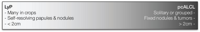
Primary cutaneous CD30+ lymphoproliferative disorders encompass lymphomatoid papulosis (LyP), primary cutaneous anaplastic large cell lymphoma (pcALCL), and indeterminate cases. LyP is a benign disorder characterized by recurrent crops of red or violaceous papulonodules. Patients with LyP are at an increased risk of a secondary malignancy. pcALCL is characterized by a solitary red to violaceous nodule or tumor larger than 20 mm. LyP is benign, is limited to the skin, and self-resolves, with a 5-year survival rate of 100%; pcALCL is limited to the skin and responsive to directed therapies, with a 5-year survival rate of over 95%. Aggressive chemotherapeutic regimens should be avoided.
Keywords: CD30(+); Cutaneous lymphoproliferative disorders; Lymphomatoid papulosis; Primary cutaneous anaplastic large cell lymphoma; Secondary cutaneous anaplastic large cell lymphoma.
Copyright © 2017 Elsevier Inc. All rights reserved.
Figures
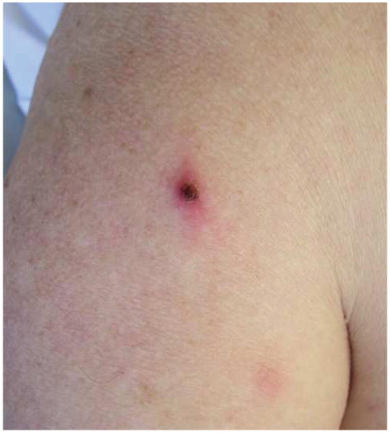
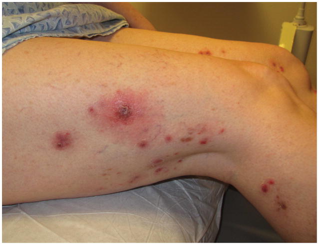
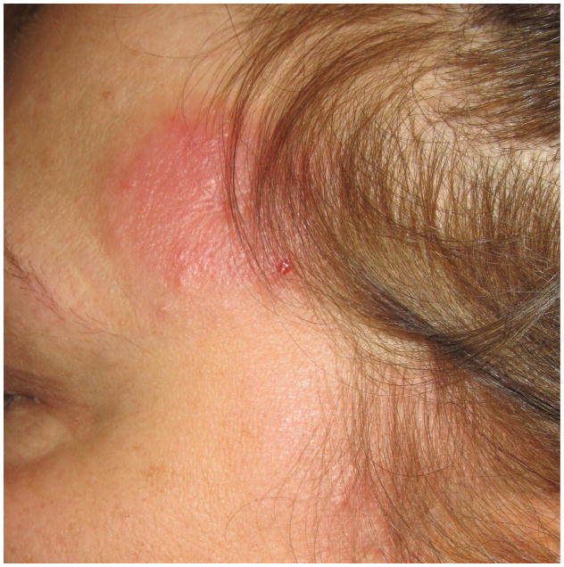
2A – Typical lesion of LyP: 6mm violaceous papule with necrotic center
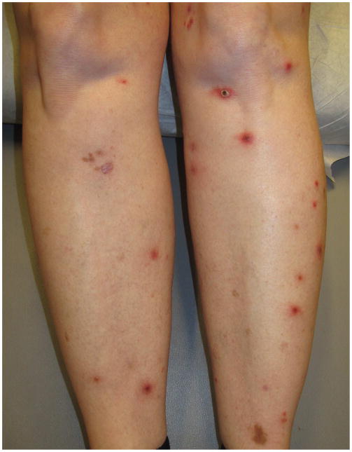
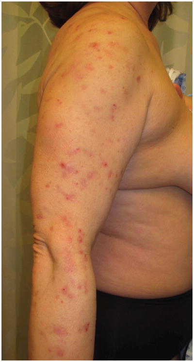
2B, C – Crops of LyP in various stages of evolution
2D – Inflamed lesion of LyP, with a surrounding crop of more typical lesions
2A – Typical lesion of LyP: 6mm violaceous papule with necrotic center
2B, C – Crops of LyP in various stages of evolution
2D – Inflamed lesion of LyP, with a surrounding crop of more typical lesions
2A – Typical lesion of LyP: 6mm violaceous papule with necrotic center
2B, C – Crops of LyP in various stages of evolution
2D – Inflamed lesion of LyP, with a surrounding crop of more typical lesions
2A – Typical lesion of LyP: 6mm violaceous papule with necrotic center
2B, C – Crops of LyP in various stages of evolution
2D – Inflamed lesion of LyP, with a surrounding crop of more typical lesions
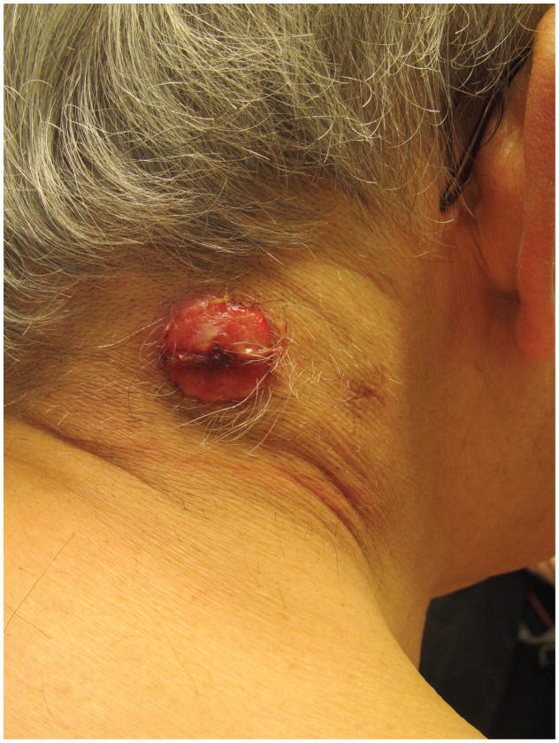
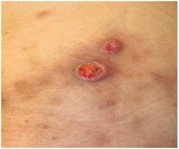
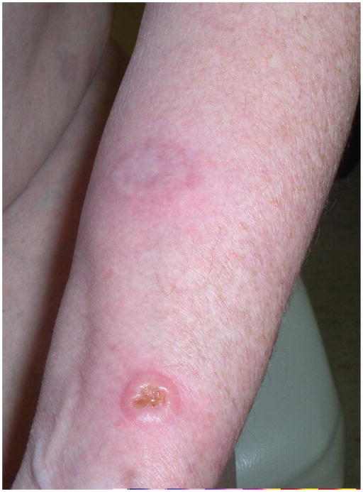
4A – Typical pcALCL tumour: Red, friable 2.5cm tumor, well-defined with central crusting
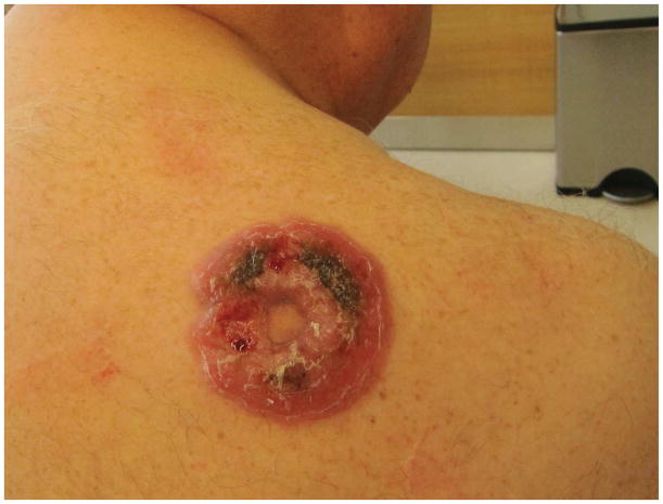
4B -- pcALCL tumor measuring 5.5 x 7.2cm with central clearing and hemorrhagic crust
4C – Early pink plaque of pcALCL
4D – Multifocal, localized and ulcerative pcALCL
4A – Typical pcALCL tumour: Red, friable 2.5cm tumor, well-defined with central crusting
4B -- pcALCL tumor measuring 5.5 x 7.2cm with central clearing and hemorrhagic crust
4C – Early pink plaque of pcALCL
4D – Multifocal, localized and ulcerative pcALCL
4A – Typical pcALCL tumour: Red, friable 2.5cm tumor, well-defined with central crusting
4B -- pcALCL tumor measuring 5.5 x 7.2cm with central clearing and hemorrhagic crust
4C – Early pink plaque of pcALCL
4D – Multifocal, localized and ulcerative pcALCL
4A – Typical pcALCL tumour: Red, friable 2.5cm tumor, well-defined with central crusting
4B -- pcALCL tumor measuring 5.5 x 7.2cm with central clearing and hemorrhagic crust
4C – Early pink plaque of pcALCL
4D – Multifocal, localized and ulcerative pcALCL
References
-
- Gilfillan MC, Noel PJ, Podack ER, Reiner SL, Thompson CB. Expression of the costimulatory receptor CD30 is regulated by both CD28 and cytokines. J Immunol. 1998;160(5):2180–7. - PubMed
-
- Smith CA, Gruss HJ, Davis T, Anderson D, Farrah T, Baker E, Sutherland GR, Brannan CI, Copeland NG, Jenkins NA, et al. CD30 antigen, a marker for Hodgkin's lymphoma, is a receptor whose ligand defines an emerging family of cytokines with homology to TNF. Cell. 1993 Jul 2;73(7):1349–60. - PubMed
-
- Bowen MA, Lee RK, Miragliotta G, Nam SY, Podack ER. Structure and expression of murine CD30 and its role in cytokine production. J Immunol. 1996 Jan 15;156(2):442–9. - PubMed
-
- Younes A, Consoli U, Snell V, Clodi K, Kliche KO, Palmer JL, Gruss HJ, Armitage R, Thomas EK, Cabanillas F, Andreeff M. CD30 ligand in lymphoma patients with CD30+ tumors. J Clin Oncol. 1997 Nov;15(11):3355–62. - PubMed
Publication types
MeSH terms
Grants and funding
LinkOut - more resources
Full Text Sources
Other Literature Sources
Medical
