

Damage-associated molecular patterns (DAMPs) released after burn are associated with inflammation and monocyte activation
- PMID: 28341255
- PMCID: PMC5373089
- DOI: 10.1016/j.burns.2016.10.001
Damage-associated molecular patterns (DAMPs) released after burn are associated with inflammation and monocyte activation
Abstract
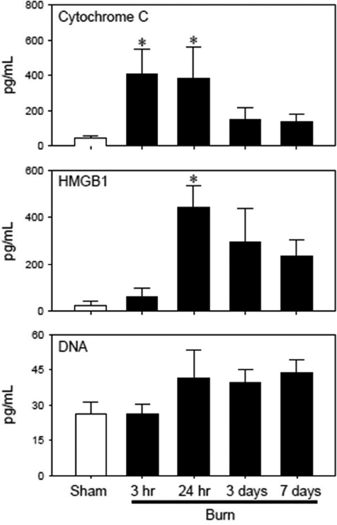
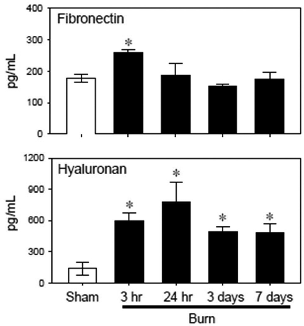
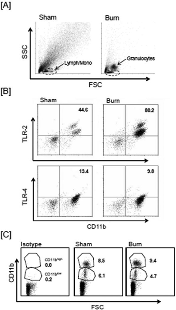
Burns are associated with activation of the innate immunity that can contribute to complications. Damage-associated molecular patterns (DAMPs) released after tissue injury play a critical role in the activation of the innate immunity, which appears to be mediated via toll-like receptors (TLRs). Previous findings have shown that TLRs and TLR-mediated responses are up-regulated after burn. Nonetheless, it is unclear what impact burn has on circulating levels of DAMPs. To study this, male C57BL/6 mice were subjected to a major burn or sham procedure. Three hours to 7days thereafter, plasma was collected and assayed for the representative DAMPs (i.e., HMGB1, cytochrome C, DNA and S100A) and extracellular cleavage products (fibronectin and hyaluronan). HMGB1, cytochrome C, fibronectin and hyaluronan levels were elevated in a time-dependent manner after burn as compared to sham levels. A significant elevation in TNF-α, IL-6 and IL-10 cytokine plasma levels was also found after burn. All cytokine levels were increased as early as 3h and remained elevated up to 24h. Circulating CD11b+ monocytes were increased at 24h after burn and showed increased expression of TLR-2. In conclusion, these findings support the concept that burn-induced elevations in circulating DAMPs are in part responsible for monocyte activation and the development of inflammatory complications under such conditions and warrants further investigation.
Keywords: Alarmins; CD11b; Danger Theory; HMGB-1; Toll-like receptors; Trauma.
Copyright © 2016 Elsevier Ltd and ISBI. All rights reserved.
Conflict of interest statement
The authors have no conflicts of interest.
Figures

References
-
- Baue AE, Durham R, Faist E. Systemic inflammatory response syndrome (SIRS), multiple organ dysfunction syndrome (MODS), multiple organ failure (MOF): are we winning the battle? Shock. 1998;10(2):79–89. - PubMed
-
- Flohe SB, Flohe S, Schade FU. Invited review: deterioration of the immune system after trauma: signals and cellular mechanisms. Innate Immun. 2008;14(6):333–344. - PubMed
-
- Krysko DV, Agostinis P, Krysko O, Garg AD, Bachert C, Lambrecht BN, et al. Emerging role of damage-associated molecular patterns derived from mitochondria in inflammation. Trends Immunol. 2011;32(4):157–164. - PubMed
MeSH terms
Substances
Grants and funding
LinkOut - more resources
Full Text Sources
Other Literature Sources
Medical
Research Materials
Miscellaneous
