Review
. 2017 Jul:133:12-30.
doi: 10.1016/j.brainresbull.2017.03.009. Epub 2017 Mar 21.

Metabolic Dysfunction in Parkinson's Disease: Bioenergetics, Redox Homeostasis and Central Carbon Metabolism

Affiliations
Review

Metabolic Dysfunction in Parkinson's Disease: Bioenergetics, Redox Homeostasis and Central Carbon Metabolism

Annadurai Anandhan et al. Brain Res Bull. 2017 Jul.

Abstract

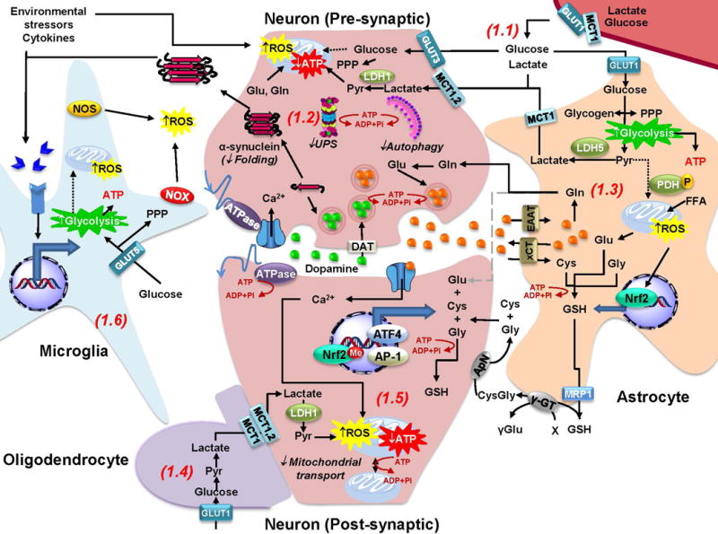
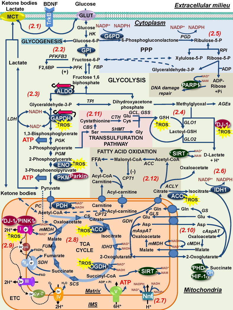
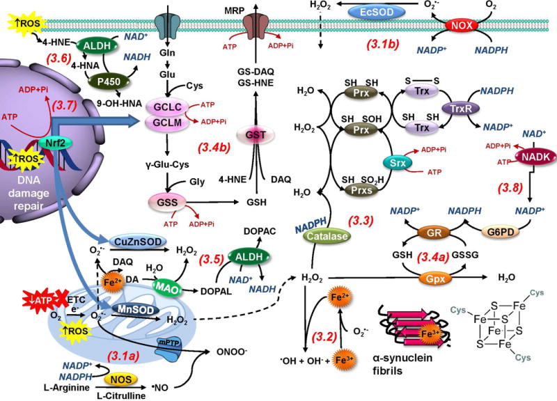
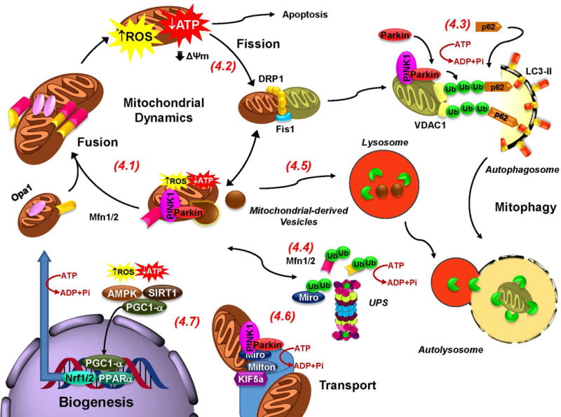
The loss of dopaminergic neurons in the substantia nigra pars compacta (SNpc) and the accumulation of protein inclusions (Lewy bodies) are the pathological hallmarks of Parkinson's disease (PD). PD is triggered by genetic alterations, environmental/occupational exposures and aging. However, the exact molecular mechanisms linking these PD risk factors to neuronal dysfunction are still unclear. Alterations in redox homeostasis and bioenergetics (energy failure) are thought to be central components of neurodegeneration that contribute to the impairment of important homeostatic processes in dopaminergic cells such as protein quality control mechanisms, neurotransmitter release/metabolism, axonal transport of vesicles and cell survival. Importantly, both bioenergetics and redox homeostasis are coupled to neuro-glial central carbon metabolism. We and others have recently established a link between the alterations in central carbon metabolism induced by PD risk factors, redox homeostasis and bioenergetics and their contribution to the survival/death of dopaminergic cells. In this review, we focus on the link between metabolic dysfunction, energy failure and redox imbalance in PD, making an emphasis in the contribution of central carbon (glucose) metabolism. The evidence summarized here strongly supports the consideration of PD as a disorder of cell metabolism.

Keywords: Bioenergetics; Glucose; Glycolysis; Mitochondria; Neurodegeneration; Oxidative stress; TCA cycle.


Figures

Figure 1
Figure 1. Redox homeostasis and metabolic coupling between neurons and glia
Redox and metabolic homeostasis is carried out by a complex interaction between neurons, glia and the extracellular microenvironment. 1.1. Glucose and lactate from blood circulation cross the blood brain barrier (BBB) through the GLUT1 (glucose transporter 1) and MCT1 (monocarboxylate transporter 1) transporters. Lactate is mainly taken up by neurons, while glucose is thought to be preferentially consumed by glial cells. 1.2 Neuronal ATP generation is dependent on oxidative phosphorylation, while glucose metabolism is primarily directed towards the PPP to generate NADPH required for antioxidant defense. Energy failure in PD is expected to impair a number of ATP-dependent processes that include: a) protein quality control mechanisms, including protein folding and protein aggregate (α-synuclein) degradation via the UPS and autophagy; b) transport of mitochondria across the axon and dendritic terminals (See 1.5); c) dopamine and glutamate capture into vesicles; and d) maintenance of ionic gradients and plasma membrane potential during synaptic transmission. 1.3. Glycolysis in astrocytes exceeds energy demands and thus lactate is shuttled as an energy substrate to neurons, which have a limited ability to upregulate glycolysis in response to mitochondrial dysfunction as it occurs in PD. Glucose flux to the TCA cycle is reduced in astrocytes due to phosphorylation of PDH. Astrocytes are able to store glucose in the form of glycogen. Glutamate (Glu) uptake from the inter-synaptic space via EAATs prevents excytotoxicity and facilitates neurotransmitter recycling via the synthesis of glutamine (Gln) that is also shuttled to neurons. In addition, Glu exchange for Cystine (that is reduced to Cysteine [Cys] inside the cell) via xCT (or Xc-), maintains Cys supply for de novo GSH synthesis. Furthermore, Nrf2 activation in response to oxidative stress promotes GSH synthesis, which is also released to provide precursors for its de novo synthesis in neurons (See 1.5). Astrocytes also have the capacity to use oxidize FFA to fuel mitochondria but its functional relevance is unclear. 1.4. Oligodendrocytes also shuttle lactate as energy fuel to myelinated axons. 1.5. In neurons, Nrf2 is repressed by methylation. Nevertheless, upregulation of antioxidant defenses is dependent on neuronal activity and the activation of ATF4 and AP-1 transcription factors. 1.6. Environmental toxicants, α-synuclein oligomers, and cytokines activate microglia and induce oxidative stress. Importantly the proinflammatory M1 phenotype of activated microglia has been reported to be associated to a switch in their metabolism from oxidative phosphorylation to glycolysis that also enhances carbon flux to the PPP.
Figure 2
Figure 2. Interaction of Parkinson disease-related genes and risk factors with bioenergetics and central carbon metabolism
2.1. BDNF released by glial cells (and activation of the tropomyosin receptor kinase B [TrkB]) has protective effects in dopaminergic cells that can potentially be associated with the regulation of neuronal bioenergetics. 2.2 In astrocytes, energy can be stored as glycogen or it can be metabolized via glycolysis to meet energy demands. Glucose metabolism is also directed to lactate production that can be shuttled to neurons. In neurons PFKFB3 is constantly degraded and thus, they depend primarily in oxidative phosphorylation to meet their energy demands. GPI has been demonstrated to exert a protective effect against α-synuclein toxicity via glycolysis. 2.3. Downstream the glycolytic pathway a number of enzymes have been shown to be altered / regulated in PD and by PD-related risk factors. Aldolase (ALDO), GAPDH and enolase (ENO) are found aggregated and oxidized in PD. Amyloid-like α-synuclein fibrils are expected to interact with and likely inhibit metastable glycolytic enzymes such as ALDO. Other groups have reported on the role of Parkin inhibiting PKM activity by ubiquitination. 2.4. Spontaneous generation of methylglyoxal is thought to account for 0.1–0.4% of glycolytic flux. Accumulation of AGEs and dopamine-related methylglyoxal derivatives is linked to PD. DJ-1 is a cofactor-independent GLO III system that detoxifies methylglyoxal while also generating D-lactate that can contribute to the maintenance of mitochondrial function. DJ-1 is a redox-sensitive protein whose protective effects against PD-related insults can be impaired by oxidative stress. 2.5 In neurons, glucose is primarily metabolized via the PPP to generate NADPH that provides reducing equivalents for antioxidant defense. Astrocytes have higher levels of NADPH and G6PD, and in glial cells, NADPH also has a pro-oxidant role by providing reducing equivalents for the generation of ROS by NOX and NOS. In addition, the ribose-5-P from the PPP is used for the generation ADP-ribose that is used during DNA-damage repair as a substrate for PARP-1 mediated ADP-ribosylation. NADPH is also regenerated by IDH1 in cytosol (2.6) and in the mitochondria by Nnt (2.7), and both systems have been reported to protect against PD-related insults MPP+ and paraquat. 2.8. Alterations in the TCA cycle have also been found associated with PD. In the mitochondria, pyruvate decarboxylation by PDH is a necessary step for the generation of acetyl-CoA and Mn (manganese), a PD-risk factor has been reported to inhibit its activity. Aconitase (ACO) inactivation by oxidative stress is a biomarker of oxidative damage induced by PD-related insults or PINK1 mutants, while a decrease in OGDH is found in PD. 2.9. A decrease in Complex I activity is found in PD. In addition Complex I has been reported to be targeted by environmental toxicants acting as Complex I inhibitors or inducing oxidative stress. PD-related genes DJ-1 and PINK1 regulate Complex I activity via direct interaction or phosphorylation of its subunits. 2.10. The mitochondrial shuttles enable electrons and precursors transport across the inner membrane. Glutamate is metabolized to glutamine by GS an enzyme exclusively present in the astrocytes. Glutamine is shuttled from astrocytes to neurons where it is metabolized back to glutamate by GLS. A number of metabolites act as substrates for the activity of signaling proteins (highlighted in green circles). Prolyl hydroxylase (PDH)-dependent hydroxylation of HIF-1α requires 2-oxoglutarate. 2.11. One-carbon metabolism of serine to glycine and cysteine (via the transsulfuration pathway) contribute to the formation of GSH. 2.12. Neurons very poorly metabolize FFA to obtain energy due the high O2 demand and resultant generation of superoxide anion (O2•−) from β-oxidation. However, 20% of total adult brain energy comes from FFA oxidation, mostly in astrocytes, but its functional relevance is unclear. Enzymes highlighted in blue circles are those within central carbon metabolism found to be modulated by oxidative stress and PD-related risk factors. PD-related genes are highlighted in red circles. Abbreviations and enzyme commission (EC) numbers for enzymes involved in central carbon metabolism: ACC, Acetyl-CoA carboxylase [EC: 6.4.1.2]; ACLY, ATP-citrate synthase [EC:2.3.3.8]; ACO, Aconitase [EC:4.2.1.3]; ALDO (A/B), Fructose-bisphosphate aldolase [EC:4.1.2.13]; cAspAT, Aspartate aminotransferase, cytoplasmic [EC:2.6.1.1 2.6.1.3]; mAspAT, Aspartate aminotransferase, mitochondrial [EC:2.6.1.1 2.6.1.7]; AR, aldose reductase [1.1.1.21]; CBS, Cystathionine beta-synthase [EC: 4.2.1.22]; CPT1, carnitine O-palmitoyltransferase 1 (CPT1 or 2 (CPT2) [EC: 2.3.1.21]; CS, Citrate synthase [EC:2.3.3.1]; CTH, Cystathionine gamma-lyase [EC: 4.4.1.1]; ENO, Enolase [EC:4.2.1.11]; FBP, fructose-1,6-bisphosphatase I [EC:3.1.3.11]; FUM, Fumarate hydratase [EC:4.2.1.2]; G3PP; Glucose-3-phosphate permease [EC: ]; G6PD, Glucose-6-phosphate 1-dehydrogenase [EC:1.1.1.49]; GAPDH, Glyceraldehyde 3-phosphate dehydrogenase [EC:1.2.1.12]; GCL, Glutamate cysteine ligase [EC: 6.3.2.2]; GDH, Glutamate dehydrogenase, [EC: 1.4.1.3]; GLO1, Glyoxalase 1 or Lactoylglutathione lyase [EC:4.4.1.5]; GLO2, Glyoxalase 2 or Hydroxyacylglutathione hydrolase [EC 3.1.2.6]; GLS, Glutaminase [EC: 3.5.1.2]; GPI, Glucose-6-phosphate isomerase [EC:5.3.1.9]; GS, Glutamine synthetase [EC:6.3.1.2]; GSS, glutathione synthetase [EC: 6.3.2.3]; HK, hexokinase [EC:2.7.1.1]; IDH1, Isocitrate dehydrogenase 1 (NADP+), soluble [EC:1.1.1.42]; IDH3, isocitrate dehydrogenase 3 (NAD+); LDH, L-lactate dehydrogenase [EC:1.1.1.27]; MDH, Malate dehydrogenase [EC:1.1.1.37]; OGDH, 2-Oxoglutarate dehydrogenase, [EC:1.2.4.2]; SCS, Succinyl-CoA synthetase [EC:6.2.1.4 6.2.1.5]; PC, Pyruvate carboxylase [EC:6.4.1.1]; PGD, 6-phosphogluconate dehydrogenase [EC:1.1.1.44]; PDH, Pyruvate dehydrogenase [EC:1.2.4.1]; PGK, phosphoglycerate kinase [EC:2.7.2.3]; PGM, Phosphoglucomutase [EC:5.4.2.2]; PFK, 6-phosphofructokinase 1 [EC:2.7.1.11]; PFKFB3, 6-phosphofucto-2-kinase/fructose-2,6-biphosphatase 3 [EC:3.1.3.46]; PKM, Pyruvate Kinase [EC:2.7.1.40]; RPI, Ribose 5-phosphate isomerase A [EC:5.3.1.6]; SDH, Succinate dehydrogenase [EC:1.3.5.1]; SHMT, Serine hydroxymethyltransferase [EC 2.1.2.1]; TPI, triose-phosphate isomerase [EC:5.3.1.1].
Figure 3
Figure 3. Bioenergetic requirements of antioxidant systems
Energy failure and ROS are the main consequences associated with mitochondrial dysfunction in PD. 3.1a. Electron leakage from the mitochondria leads to one electron reduction of O2 to O2•− that can be dismutated by SODs to H2O2.NO can outcompete SODs reacting with O2•− to generate ONOO. 3.1b. In the plasma membrane O2•− can be generated by NOXs. 3.2. In the presence of metals such as Fe, H2O2 and O2•− generate OH through the Fenton/Haber-Weiss reaction. In PD, α-synuclein, neuromelanin and Fe-S clusters are important pools of Fe. 3.3. Catalase and Prxs catalyze H2O2 degradation. The Trx/TrxR system supplies reducing equivalents for most Prxs. Prx hyperoxidation is reversed by Srx. 3.4a. Gpxs detoxifies peroxides using GSH which is reduced back from GSSG by GR using NADPH. 3.4b. GSH also detoxifies electrophiles (DAQ and 4-HNE) via the action of GSTs, and these adducts can be transported outside of the cell to become eliminated by the activity of MRP proteins. 3.5. In the cytosol, dopamine (DA) becomes auto-oxidized in the presence of Fe generating DAQs and ROS. DA metabolism by MAO also generates ROS and DOPAL, which is further metabolized by ALDHs. 3.6. ALDHs also detoxify 4-HNE into 4-hydroxy-2-nonenoic acid (HNA) that is metabolized by cytochrome P450 enzymes (P450). 3.7. Oxidative stress triggers antioxidant response elements (AREs)-regulated gene transcription by Nrf2, which promotes GCLC/GCLM, CuZnSOD and MnSOD transcription. 3.8. A number of antioxidant systems and ROS generating enzymes utilize reducing equivalents from NADPH (in blue letters) that is regenerated by enzymes such as G6PD, and synthesized de novo via the phosphorylation of NAD+ by NAD-kinase (NADK). Thus, antioxidant systems are tightly coupled to NAD+ and central carbon metabolism. In addition, many antioxidant enzymes and stress responses (DNA-damage repair and the transcription of antioxidant enzymes by Nrf2) require energy consumption (ATP, highlighted in dark red) demonstrating that redox homeostasis is tightly coupled to bioenergetics and cell metabolism.
Figure 4
Figure 4. Mitochondrial dynamics and biogenesis are coupled to oxidative stress and energy failure
4.1. Mitochondrial fusion rescues “moderately” dysfunctional mitochondria by enabling their “damaged” content to be mixed between neighboring mitochondria. Fusion is impaired by the ubiquitination of Mfns by PINK1 and Parkin (See 4.4). 4.2. Fission, transforms damaged mitochondria into a form suitable for engulfment by mitophagy, and it has been suggested to facilitate transport of mitochondria into terminals (See 4.6). 4.3. Parkin translocates to the mitochondria and interacts with PINK1 in response to a loss in membrane potential (ΔΨm) induced by energy failure or oxidative damage. VDAC1 is polyubiquitinated and recruits p62, which in turn interacts with LC3 at the autophagosomal membrane. Finally, engulfed damaged mitochondria are degraded after the autophagosome fuses with the lysosome and lysosomal hydrolases are released within the fused autolysosome. 4.5. It has also been proposed that Parkin and PINK1 induce the formation of mitochondrial derived vesicles that translocate damaged mitochondrial proteins into lysosomes. 4.6. Transportation of mitochondria to axonal and dendritic terminations is essential to meet energy demands associated with synaptic transmission. PINK1 and Parkin mediate phosphorylation, ubiquitination and degradation of Miro leading to the detachment of mitochondria from microtubules. 4.7. Oxidative stress and energy failure activate PGC-1α to stimulate mitochondrial biogenesis. Energy failure in PD is expected to impair transport of mitochondria into terminals, and the mitochondrial quality control mechanisms mitophagy and UPS. 4.4. The proteasome has also been proposed to translocate to the mitochondria and mediate the degradation of damaged proteins. (ATP consumption is highlighted in red).

References

    1. Effects of tocopherol and deprenyl on the progression of disability in early Parkinson’s disease. N Engl J Med. 1993;328:176–183. - PubMed
    1. Abdin AA, Hamouda HE. Mechanism of the neuroprotective role of coenzyme Q10 with or without L-dopa in rotenone-induced parkinsonism. Neuropharmacology. 2008;55:1340–1346. - PubMed
    1. Abou-Sleiman PM, Muqit MM, Wood NW. Expanding insights of mitochondrial dysfunction in Parkinson’s disease. Nat Rev Neurosci. 2006;7:207–219. - PubMed
    1. Abramov AY, Gegg M, Grunewald A, Wood NW, Klein C, Schapira AH. Bioenergetic consequences of PINK1 mutations in Parkinson disease. PLoS One. 2011;6:e25622. - PMC - PubMed
    1. Ahmed SS, Santosh W, Kumar S, Christlet HT. Metabolic profiling of Parkinson’s disease: evidence of biomarker from gene expression analysis and rapid neural network detection. J Biomed Sci. 2009;16:63. - PMC - PubMed