. 2017 Jun;23(6):847-859.
doi: 10.1261/rna.060749.117. Epub 2017 Mar 24.

Structural landscape of base pairs containing post-transcriptional modifications in RNA

Affiliations

Structural landscape of base pairs containing post-transcriptional modifications in RNA

Preethi P Seelam et al. RNA. 2017 Jun.

Abstract

Base pairs involving post-transcriptionally modified nucleobases are believed to play important roles in a wide variety of functional RNAs. Here we present our attempts toward understanding the structural and functional role of naturally occurring modified base pairs using a combination of X-ray crystal structure database analysis, sequence analysis, and advanced quantum chemical methods. Our bioinformatics analysis reveals that despite their presence in all major secondary structural elements, modified base pairs are most prevalent in tRNA crystal structures and most commonly involve guanine or uridine modifications. Further, analysis of tRNA sequences reveals additional examples of modified base pairs at structurally conserved tRNA regions and highlights the conservation patterns of these base pairs in three domains of life. Comparison of structures and binding energies of modified base pairs with their unmodified counterparts, using quantum chemical methods, allowed us to classify the base modifications in terms of the nature of their electronic structure effects on base-pairing. Analysis of specific structural contexts of modified base pairs in RNA crystal structures revealed several interesting scenarios, including those at the tRNA:rRNA interface, antibiotic-binding sites on the ribosome, and the three-way junctions within tRNA. These scenarios, when analyzed in the context of available experimental data, allowed us to correlate the occurrence and strength of modified base pairs with their specific functional roles. Overall, our study highlights the structural importance of modified base pairs in RNA and points toward the need for greater appreciation of the role of modified bases and their interactions, in the context of many biological processes involving RNA.

Keywords: X-ray crystal structures; base-pair parameters; interaction energies; modified base pairs; post-transcriptional modifications.


Figures

FIGURE 1.
FIGURE 1.
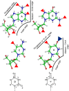
Schematic representation of modified base pairs showing their interacting edges. Red triangles represent modification involving methyl group substitution, whereas the blue triangle represents substitution of oxygen with sulfur atom. The ribose sugar is represented by r in the structures of dihydrouridine (D) and pseudouridine (ψ).
FIGURE 2.
FIGURE 2.
(A) Schematic representation of cis (C) or trans (T) orientation of the glycosidic bond. (B) List of 12 RNA base-pairing families. W, H, and S represent Watson–Crick, Hoogsteen, and sugar edges, respectively.
FIGURE 3.
FIGURE 3.
(A) Percent distribution of total 207 crystal structures in the data set as a function of RNA type. (B) Percent distribution of those 135 crystal structures as a function of RNA type that contain at least one modified base.
FIGURE 4.
FIGURE 4.
Schematic representation of most commonly observed modified base pairs in tRNA sequences. (A) Distribution of modified base pairs in tRNA sequences divided according to the domains of life. (BK) Presence of modified base pairs in 10 major base pair positions (represented by red circles) in tRNA structures. The newly identified modified base pair combinations observed from sequence analysis are shown in bold in the corresponding tables.
FIGURE 5.
FIGURE 5.
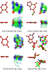
Structural alignment of crystal occurrences of modified base pairs (with occurrence frequency ≥30) with their corresponding optimized structures. Occurrence frequency and average RMSD (in Å) with respect to the optimized structure (ball and stick, red) is given in the parentheses.
FIGURE 6.
FIGURE 6.
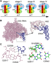
(A) Flexible 3′-CCA end (white box) of tRNA during various stages of tRNA accommodation at the A-site (yellow box) of the 70S ribosome. The neighboring P-site of rRNA is shown as a red box. (B) Interaction of 3′-CCA containing amino acceptor arm of tRNA blue) of tRNA (blue) with the A-loop (H92) of 23S rRNA (pink). (C) Structure of base quartet formed from the interaction of the preformed G-minor base triplet (C2542:G2617:Gm2588) present at H92 of rRNA and the C75 of the 3′-CCA of tRNA. Alignment of the preformed rRNA triplet containing the 2′-methylated G2588 present in the crystal structure of the tRNA:rRNA complex of H. marismortui (PDB: 3cme), with the corresponding triplet containing the unmodified G2588 present in one of the crystal structures of the tRNA:rRNA complex of E. coli (PDB: 4v9d).
FIGURE 7.
FIGURE 7.
Presence of modified base pairs at the binding site of antibiotics streptomycin and paromomycin. (A) Structure of 16S rRNA bound to streptomycin (red) and paromomycin (orange). (B,C) Antibiotic-binding pocket with surrounding proteins (S12). (D,E) Interaction of base pairs C522:527 and C1407:G1494 present in the binding pocket with the antiobiotics streptomycin and paromomycin, respectively. (F) Hydrophobic cloud created by surrounding amino acid residues around the methyl group attached to ring II of streptomycin (red). Methyl modification of G527 or C1407 at the nucleobase sites represented by blue circles result in resistance to antibiotic binding.
FIGURE 8.
FIGURE 8.
Modified base pairs involved in higher-order interaction motifs.

Similar articles

Cited by

References

    1. Ahlrichs R, Elliott SD, Huniar U. 1998. Quantum chemistry: large molecules–small computers. Ber Bunsen-Ges Phys Chem 102: 795–804.
    1. Altschul SF, Gish W, Miller W, Myers EW, Lipman DJ. 1990. Basic local alignment search tool. J Mol Biol 215: 403–410. - PubMed
    1. Bansal M, Bhattacharyya D, Ravi B. 1995. NUPARM and NUCGEN: software for analysis and generation of sequence dependent nucleic acid structures. Comput Appl Biosci 11: 281–287. - PubMed
    1. Becke AD. 1993. Density-functional thermochemistry. III. The role of exact exchange. J Chem Phys 98: 5648–5652.
    1. Bhattacharya S, Mittal S, Panigrahi S, Sharma P, Seelam PP, Paul R, Halder S, Halder A, Bhattacharyya D, Mitra A. 2015. RNABP COGEST: a resource for investigating functional RNAs. Database 2015: bav011. - PMC - PubMed

Publication types

LinkOut - more resources