. 2017 May 19;292(20):8223-8235.
doi: 10.1074/jbc.M116.762609. Epub 2017 Mar 24.

Adductome-based identification of biomarkers for lipid peroxidation

Affiliations

Adductome-based identification of biomarkers for lipid peroxidation

Takahiro Shibata et al. J Biol Chem. .

Abstract

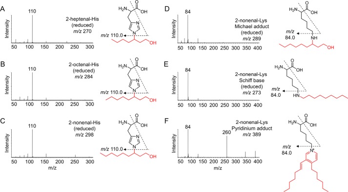
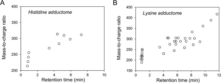
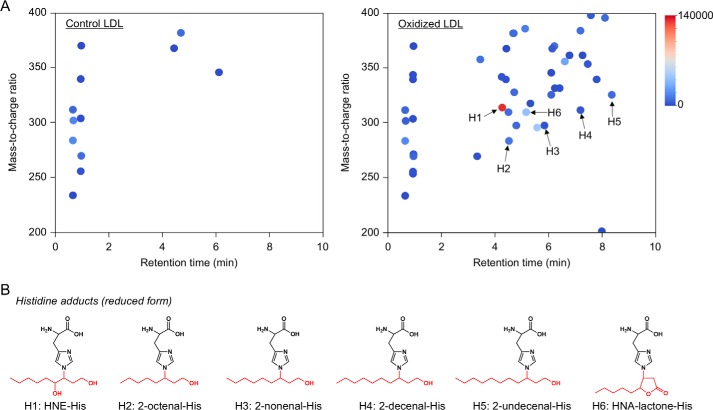
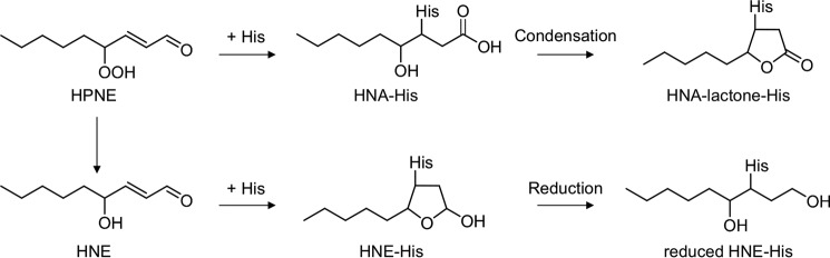
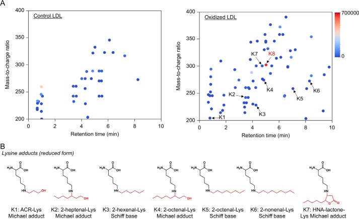
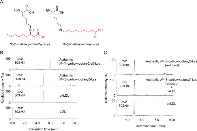
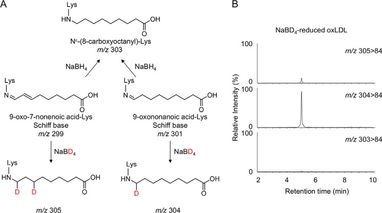
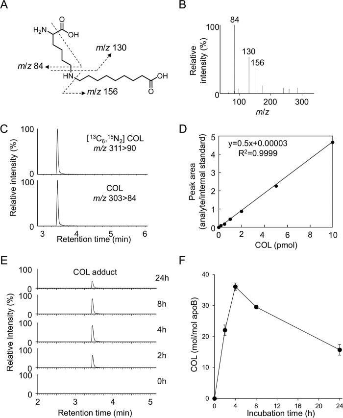
Lipid peroxidation is an endogenous source of aldehydes that gives rise to covalent modification of proteins in various pathophysiological states. In this study, a strategy for the comprehensive detection and comparison of adducts was applied to find a biomarker for lipid peroxidation-modified proteins in vivo This adductome approach utilized liquid chromatography with electrospray ionization tandem mass spectrometry (LC-ESI-MS/MS) methods designed to detect the specific product ions from positively ionized adducts in a selected reaction monitoring mode. Using this procedure, we comprehensively analyzed lysine and histidine adducts generated in the in vitro oxidized low-density lipoproteins (LDL) and observed a prominent increase in several adducts, including a major lysine adduct. Based on the high resolution ESI-MS of the adduct and on the LC-ESI-MS/MS analysis of the synthetic adduct candidates, the major lysine adduct detected in the oxidized LDL was identified as Nϵ-(8-carboxyoctanyl)lysine (COL). Strikingly, a significantly higher amount of COL was detected in the sera from atherosclerosis-prone mice and from patients with hyperlipidemia compared with the controls. These data not only offer structural insights into protein modification by lipid peroxidation products but also provide a platform for the discovery of biomarkers for human diseases.

Keywords: aldehyde; biomarker; lipid oxidation; lipoprotein; mass spectrometry (MS); protein chemical modification.


Conflict of interest statement

The authors declare that they have no conflicts of interest with the contents of this article

Figures

Figure 1.
Figure 1.
Strategy of histidine and lysine adductome analysis.
Figure 2.
Figure 2.
Collision-induced dissociation of the [M + H]+ of histidine and lysine adducts at the collision energy of 25 V and proposed structures of individual ions. A, reduced 2-heptenal-His (m/z 270). B, reduced 2-octenal-His (m/z 284). C, reduced 2-nonenal-His (m/z 298). D, reduced 2-nonenal-Lys Michael adduct (m/z 289). E, reduced 2-nonenal-Lys Schiff base (m/z 273). F, 2-nonenal-Lys pyridinium adduct (m/z 389).
Figure 3.
Figure 3.
Adductome maps of known histidine adducts (A) and lysine adducts (B). The specific product ions from positively ionized histidine and lysine adducts were analyzed by LC-ESI-MS/MS in the SRM mode transmitting the [M + H]+ >110 (for histidine adduct, A) or [M + H]+ >84 (for lysine adduct, B) transition.
Figure 4.
Figure 4.
Adductome analysis of histidine adducts detected in the oxidized LDL. A, adductome maps of histidine adducts detected in the control (left) or Cu2+-oxidized LDL (right). The adductome maps are shown with a color gradient encoding the relative abundance from blue (low) to red (high). The known histidine adducts are indicated by arrows labeled H1–H6. B, chemical structure of histidine adducts (H1–H6) detected in the Cu2+-oxidized LDL.
Figure 5.
Figure 5.
Proposed mechanism for the formation of HNE-His and HNA-lactone-His adducts.
Figure 6.
Figure 6.
Adductome analysis of lysine adducts detected in the oxidized LDL. A, adductome maps of lysine adducts detected in the control (left) or Cu2+-oxidized LDL (right). The adductome maps are shown with a color gradient encoding the relative abundance from blue (low) to red (high). The known lysine adducts are indicated by arrows labeled K1–K7. The adduct K8 indicates a putative adduct that was detected at a high level. B, chemical structure of lysine adducts (K1–K7) detected in the Cu2+-oxidized LDL.
Figure 7.
Figure 7.
Identification of COL adduct as a major lysine adduct in oxidized LDL. A, chemical structure of Nϵ-(1-carboxyoctan-2-yl)-lysine (left) and COL adduct (right). B, LC-ESI-MS/MS analysis of authentic COL adduct. C, co-injection experiment on the LC-ESI-MS/MS of synthetic COL adduct with the acid-hydrolyzed sample from oxidized LDL.
Figure 8.
Figure 8.
Deuterium incorporation experiments using NaBD4. A, reduction of 9-oxo-7-nonenoic acid-Lys and 9-oxononanoic acid-Lys Schiff base adducts by NaBD4. B, LC-ESI-MS/MS analysis of the NaBD4-reduced oxidized LDL. The oxidized LDL was analyzed by LC-ESI-MS/MS in the SRM mode (upper, 305 > 84; middle, 304 > 84; lower, 303 > 84) following NaBD4 reduction and acid hydrolysis.
Figure 9.
Figure 9.
Quantification of COL adduct in Cu2+-oxidized LDL. A and B, collision-induced dissociation of the [M + H]+ of COL adduct at the collision energy of 25 V. A, proposed structures of individual ions. C, LC-ESI-MS/MS analysis of [13C6,15N3]COL adduct. Upper, SRM for [13C6,15N3]COL adduct (m/z 311 > 90); lower, SRM for COL adduct (m/z 303 84). D, calibration curves for COL adduct. E and F, time-dependent formation of COL adduct in the Cu2+-oxidized LDL. The LDLs were analyzed by LC-ESI-MS/MS in the SRM mode following NaBH4 reduction and acid hydrolysis.
Figure 10.
Figure 10.
Collision-induced dissociation of the [M + H]+ of reduced HNE-histidine and HNA-lactone-histidine adducts at the collision energy of 25 V. A–D, reduced HNE-His adducts (m/z 314). E–H, HNA-lactone-His adduct (m/z 310). Proposed structures of individual ions (A and E). C, LC-ESI-MS/MS analysis of 15N3-reduced HNE-His adduct. Upper, SRM for 15N3-reduced HNE-His (m/z 317 > 113); lower, SRM for reduced HNE-His (m/z 314 > 110). D, calibration curves for reduced HNE-His adduct. G, LC-ESI-MS/MS analysis of [15N3]HNA-lactone-His adduct. Upper, SRM for [15N3]HNA-lactone-His (m/z 313 > 113); lower, SRM for reduced HNA-lactone-His (m/z 310 > 110). H, calibration curves for HNA-lactone-His adduct.
Figure 11.
Figure 11.
Quantification of histidine adducts in Cu2+-oxidized LDL. Time-dependent formation of reduced HNE-His (A and C) and HNA-lactone-His adducts (B and C) in the Cu2+-oxidized LDL. The LDLs were analyzed by LC-ESI-MS/MS in the SRM mode following NaBH4 reduction and acid hydrolysis.
Figure 12.
Figure 12.
Quantification of COL adduct in vivo. A and B, formation of COL adduct in the sera from control and spontaneously hyperlipidemic mice. *, p < 0.05. C, formation of COL adduct in lipoproteins factions from spontaneously hyperlipidemic mice. ***, p < 0.005. D, formation of COL adduct in the sera from normal subjects and patients with hyperlipidemia. **, p < 0.01. E, correlation between the levels of COL and those of LDL.

References

    1. Gutteridge J. M., and Halliwell B. (1990) The measurement and mechanism of lipid peroxidation in biological systems. Trends Biochem. Sci. 15, 129–135 - PubMed
    1. Esterbauer H., Schaur R. J., and Zollner H. (1991) Chemistry and biochemistry of 4-hydroxynonenal, malonaldehyde and related aldehydes. Free Radic. Biol. Med. 11, 81–128 - PubMed
    1. Uchida K. (2003) Histidine and lysine as targets of oxidative modification. Amino Acids 25, 249–257 - PubMed
    1. Steinberg D., Parthasarathy S., Carew T. E., Khoo J. C., and Witztum J. L. (1989) Beyond cholesterol. Modifications of low-density lipoprotein that increase its atherogenicity. N. Engl. J. Med. 320, 915–924 - PubMed
    1. Steinberg D. (1995) Role of oxidized LDL and antioxidants in atherosclerosis. Adv. Exp. Med. Biol. 369, 39–48 - PubMed