. 2018 May;67(5):847-859.
doi: 10.1136/gutjnl-2016-313214. Epub 2017 Mar 24.

Gut symbiotic microbes imprint intestinal immune cells with the innate receptor SLAMF4 which contributes to gut immune protection against enteric pathogens

Affiliations

Gut symbiotic microbes imprint intestinal immune cells with the innate receptor SLAMF4 which contributes to gut immune protection against enteric pathogens

Allison Cabinian et al. Gut. 2018 May.

Abstract

Background: Interactions between host immune cells and gut microbiota are crucial for the integrity and function of the intestine. How these interactions regulate immune cell responses in the intestine remains a major gap in the field.

Aim: We have identified the signalling lymphocyte activation molecule family member 4 (SLAMF4) as an immunomodulator of the intestinal immunity. The aim is to determine how SLAMF4 is acquired in the gut and what its contribution to intestinal immunity is.

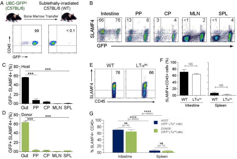
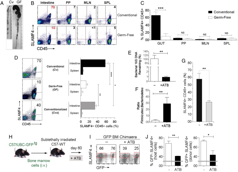
Methods: Expression of SLAMF4 was assessed in mice and humans. The mechanism of induction was studied using GFPtg bone marrow chimaera mice, lymphotoxin α and TNLG8A-deficient mice, as well as gnotobiotic mice. Role in immune protection was revealed using oral infection with Listeria monocytogenes and Cytobacter rodentium.

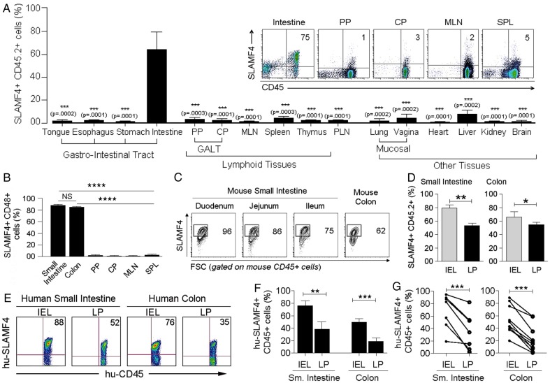
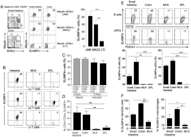
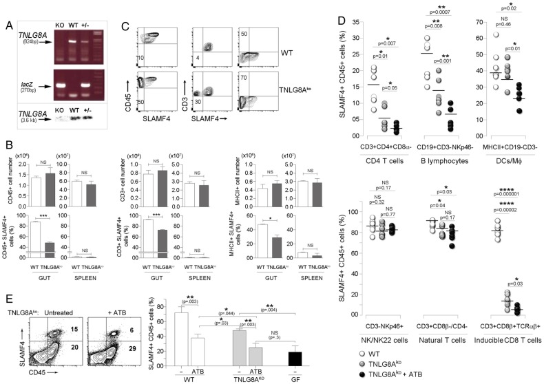
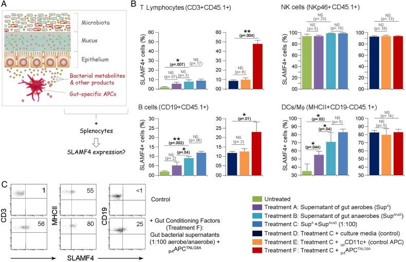
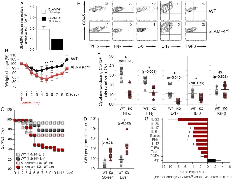
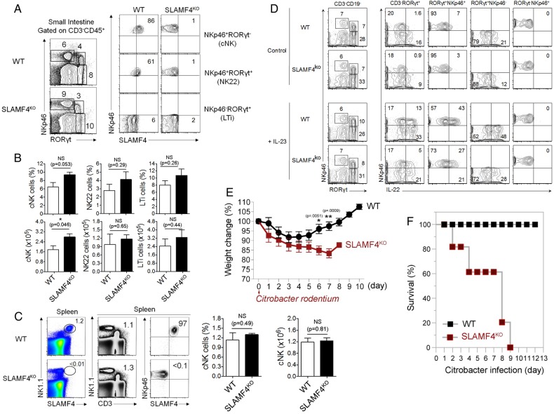
Results: SLAMF4 is a selective marker of intestinal immune cells of mice and humans. SLAMF4 induction occurs directly in the intestinal mucosa without the involvement of the gut-associated lymphoid tissue. Gut bacterial products, particularly those of gut anaerobes, and gut-resident antigen-presenting cell (APC) TNLG8A are key contributors of SLAMF4 induction in the intestine. Importantly, lack of SLAMF4 expression leads the increased susceptibility of mice to infection by oral pathogens culminating in their premature death.

Conclusions: SLAMF4 is a marker of intestinal immune cells which contributes to the protection against enteric pathogens and whose expression is dependent on the presence of the gut microbiota. This discovery provides a possible mechanism for answering the long-standing question of how the intertwining of the host and gut microbial biology regulates immune cell responses in the gut.

Keywords: ANTIBIOTICS; ENTERIC INFECTIONS; GASTROINTESTINAL IMMUNE RESPONSE; INTESTINAL BACTERIA; MUCOSAL IMMUNITY.


Conflict of interest statement

Competing interests: None declared.

Figures

Figure 1
Figure 1
Signalling lymphocyte activation molecule family member 4 (SLAMF4) is a marker of intestinal immune cells. (A) SLAMF4 expression was assessed on haematopoietic-derived (CD45+) cells in the Peyer's patches (PP) and caecal patch (CP) which belong to the gut-associated lymphoid tissue (GALT), mesenteric lymph nodes (MLN), spleen (SPL), peripheral lymph nodes (PLN), as well as other organs as indicated. Bar graph summarises SLAMF4 expression as means of % CD45 cells that are SLAMF4+. (Inset) Dot plots show SLAMF4 expression by CD45+ cells in different tissues. (B) Gut mucosal CD45+ cells express both SLAMF4 and its ligand CD48. Data are presented as means of % SLAMF4+CD45+ cells that are CD48+. Data shown in (A, B) are from ten experiments using two to three mice per experiment for intestinal tissues and four experiments using two to three mice per experiment for other organs. (C) Dot plots show SLAMF4 expression by CD45+ cells in the intraepithelial (IEL) compartment of different segments of the small intestine and colon. Numbers indicate % SLAMF4+CD45+ cells. (D) Murine IEL and lamina propria (LP) fractions were prepared from the small intestine and colon and stained for flow cytometry. Bar graphs show SLAMF4 expression as means of % CD45+ cells that are SLAMF4+ in the IEL compartment versus those in the LP. Data shown in (C, D) are from n=6. (E) SLAMF4 expression was assessed on human small intestine (n=7) and colon (n=10) samples. Dot plots show SLAMF4 expression on human CD45+ cells in the IEL and LP compartments. (F) Data are shown as means of % human CD45+ cells expressing SLAMF4 and (G) relationship between IEL and LP for each tissue sample. Error bars represent SEM. A two-tailed Student's t-test distribution with paired groups of samples was evaluated for statistical significance. A value of p>0.05 is considered not significant (NS); *p<0.05, **p<0.005, ***p<0.005, ****p<0.00005.
Figure 2
Figure 2
Signalling lymphocyte activation molecule family member 4 (SLAMF4) is expressed by different immune cell types in the intestinal mucosa. (A) Lamina propria cells were first gated on CD45+CD3− cells. Dot plots show three NK cell subtypes identified based on the expression of NKp46 and the transcriptional factor RORγt. Numbers indicate the % of each cell subset (left) as well as % SLAMF4+ cells (right). Bar graph summarises % SLAMF4+ cells gated on each subtype. Data are from four experiments using three mice per experiment. (B) SLAMF4 expression was assessed on CD8αα+, CD8αβ+TCRαβ+ and TCRγδ+ T cells in the intestine, mesenteric lymph nodes (MLN) and spleen (SPL). Numbers indicate % of SLAMF4+ cells. (C) Summary data are shown as means of % gut CD8 T cell subsets that are SLAMF4+. Data are from four experiments using two mice per experiment. (D) Bar graph shows SLAMF4 expression on CD4+ cells in the intestine versus periphery. Data are presented as means of % CD4+ (CD3+CD4+CD8α−CD8β−) cells that are SLAMF4+. Data are from three experiments using three mice per experiment. (E) SLAMF4 expression on B cells (CD45+CD3-B220+CD19+) and plasmacytoid dendritic cells (pDCs, CD45+PDCA1+CD11c+). Below, bar graphs summarise FACS data. (F) Bar graphs show SLAMF4 expression on dendritic cells (DC, CD11c+CD19−CD3−F4/80−, left) and macrophages (Mφ, CD11c−CD11b+F4/80+, right). Data are shown as means of % DCs and Mφ that are SLAMF4+. Data shown in (E, F) are from three experiments using three to four mice per experiment. Data are shown as means±SEM. Error bars represent SEM. A two-tailed Student's t-test distribution with paired groups of samples was evaluated for statistical significance. *p<0.05, **p<0.005, ***p<0.0005.
Figure 3
Figure 3
Signalling lymphocyte activation molecule family member 4 (SLAMF4) induction occurs directly in the gut mucosa and independently of the gut-associated lymphoid tissue (GALT). (A) B6.GFPtg bone marrow cells were adoptively transferred into sublethally irradiated B6 mice. Plots show GFP expression on donor (left) and host (right) CD45+ cells. Data are representative of n=10. (B) Eight weeks later, mice were euthanised to isolate cells from the gut mucosa, Peyer's patch (PP), caecal patch (CP), mesenteric lymph nodes (MLN) and spleen (SPL). Numbers indicate % of host (GFP−, top left) and donor (GFP+, top right) SLAMF4+ cells. Summary of data displayed in (B) are shown in (C) and (D), respectively. (E) Expression of SLAMF4 in the intestinal mucosa of B6.WT and B6.Ltαko mice. Data are representative of n=9. (F) Summary of data indicating SLAMF4 expression as means of % CD45+ cells in the intestine and spleen. (G) B6.GFPtg bone marrow cells were adoptively transferred into sublethally irradiated B6.Ltαko recipients as described in (A). Data are shown as means of % host (GFP−Ltα−/−) and donor (GFP+Ltα+/+) CD45.2+ cells that are SLAMF4+. Data are from three experiments using two (C,D,F) and three (g) mice per experiment. Data are shown as means±SEM. Error bars represent SEM. A two-tailed Student's t-test distribution with paired groups of samples was evaluated for statistical significance. p>0.05 is considered not significant (NS); *p<0.05, ***p<0.0005, ****p<0.00005. WT, wild type.
Figure 4
Figure 4
Signalling lymphocyte activation molecule family member 4 (SLAMF4) expression in the gut mucosa is dependent on the continued presence of the commensal microflora. (A) Representative photographs illustrating colon morphology in conventional (Cv) and germ-free (GF) BALB/c mice. (B) SLAMF4 expression on intestinal CD45+ cells is inhibited in GF animals, but there is no difference in SLAMF4 expression in Peyer's patches (PP), mesenteric lymph nodes (MLN) or spleen (SPL). (C) Summary of SLAMF4 expression on CD45+ cells shown in (B). (D) Dot plots show SLAMF4 expression on gut CD45+ cells in Cv, GF and conventionalised (Cvz) mice. Bar graph summarises data as means of % CD45+ cells that are SLAMF4+ in the intestine and spleen. Data shown in (B–D) are from three experiments using two mice per animal group per experiment. (E–G) B6 mice were left untreated (−) or treated with a combination of four antibiotics (ATB, ampicillin, metronidazole, neomycin, vancomycin) in the drinking water for 4 weeks. (E) Faecal DNA was made from intestinal contents and analysed for total bacterial contents. (F) Faecal DNA analysed for proportions (normalised to total bacteria) of Bacteroidetes and Firmicutes by qPCR. Data displayed are fold Bacteroidetes/Firmicutes. (G) Bar graph shows % gut CD45+ cells that remain SLAMF4+ after ATB treatment. Data shown in (E–G) are from five experiments using two mice per animal group per experiment. (H) B6.GFPtg bone marrow chimaera mice were left untreated or treated with ATB as described in (G). (I) Dot plots show SLAMF4 expression on intestinal cells of untreated and ATB-treated mice. Numbers indicate % SLAMF4+ cells. (J) Bar graphs summarise % CD45+ cells remaining SLAMF4+ among host (GFP−, right) and donor (GFP+, left) intestinal cells (n=5 per animal group). Overall data are shown as means±SEM. Error bars represent SEM. A two-tailed Student's t-test distribution with paired groups of samples was evaluated for statistical significance. *p<0.05, **p<0.005, ***p<0.0005, ****p<0.0001.
Figure 5
Figure 5
Role of TNLG8A in signalling lymphocyte activation molecule family member 4 (SLAMF4) induction. (A) Lack of TNLG8A gene expression in knockout mice. Amplification of two PCR products, TNLG8A (824bp) and lacZ (270bp), revealed the deletion of the TNLG8A gene in knockout mice (top), and absence of the TNLG8A product (3.6 kb) by Southern blot analysis confirmed this gene deletion (bottom). (B) Pooled IEL and LP cells from wild-type (WT) and TNLG8Ako mice were analysed by flow cytometry. Bar graphs summarise SLAMF4 expression as means of numbers (top) and % (bottom) of total leucocytes (CD45+), T cells (CD3+MHCII−) and APCs (MHCII+CD3−) that are SLAMF4+. (C) Dot plots show SLAMF4 expression in the intestinal mucosa of WT and TNLG8Ako mice. Numbers indicate % SLAMF4− cells. Data shown in (A–C) are from three experiments using three mice per animal group per experiment. (D) WT and TNLG8Ako mice were left untreated or treated with ATB as described in figure 4E. Graphs summarise % SLAMF4+ cells in the intestinal mucosa of untreated WT (control) versus untreated and ATB-treated TNLG8Ako animals. Data show significant reduction of SLAMF4 expression on CD4T cells (CD3+CD4+CD8α−), inducible CD8T cells (CD3+CD8β+TCRαβ+), B cells (CD3−CD19+NKp46−) and DC/MΦ (CD3−CD19−MHCII+), but little or no decrease on natural CD8T cells (CD3+CD8α+CD8β−CD4−) and NK/NK22 cells (CD3−CD19−NKp46+). (E) Dot plots show SLAMF4 expression on gut leucocytes in untreated and ATB-treated TNLG8Ako animals. Bar graph summarises SLAMF4 expression as means of % intestinal CD45+ cells that are SLAMF4+ in untreated (−) and ATB-treated WT and TNLG8Ako mice in comparison with germ-free (GF) animals. Data are from three experiments using two (untreated) and three (ATB-treated) mice per animal group per experiment. Data are shown as means ±SD. Error bars represent SD. A two-tailed Student's t-test distribution with paired sample groups was evaluated for statistical significance. Value of p>0.05 was considered not significant (NS); *p<0.05, **p<0.005, ***p<0.0005, ****p<0.0001.
Figure 6
Figure 6
Transfer of signalling lymphocyte activation molecule family member 4 (SLAMF4) expression on systemic immune cells. (A) The hypothesis that is being tested is that gut bacterial products and gut-specific APCs provide the environmental conditions for SLAMF4 induction in the intestine, and provision of these gut signals enables the transfer of this phenotype to peripheral immune cells. (B) T lymphocytes (CD3+), natural killer (NK) cells (NKp46+), B cells (CD19+) and other APCs (CD19-MHCII+) were magnetically purified from CD45.1 splenocytes. Subsequently, they were cultured with 8 U (normalised value of OD600×μL) of aerobe and anaerobe supernatants in the presence or absence of CD45.2+ APCs (splCD11c+ cells or gutAPCTNLG8A). Eighteen hours later, CD45.1+ cells were examined for the expression of SLAMF4 by flow cytometry. Data are shown as means of % CD45.1+ cells±SD. Error bars represent SD. Data are the summary of four experiments using two mice per treatment per experiment. (C) CD45.1 splenocyte types were treated with bacterial broths in the presence of (CD45.2+) splCD11c+ cells (control, upper) or with gut bacterial supernatants (1:100 anaerobe/aerobe) in the presence of gutAPCTNLG8 (bottom). Dot plots show expression of SLAMF4 on T (CD3+) cells, B (CD19+) cells and other APCs (CD19-MHCII+). Numbers indicate % SLAMF4+ cells. A two-tailed Student's t-test distribution with paired sample groups was evaluated for statistical significance. A value of p>0.05 was considered not significant (NS); *p<0.05, ***p<0.0005.
Figure 7
Figure 7
Lack of signalling lymphocyte activation molecule family member 4 (SLAMF4) renders mice susceptible to Listeria monocytogenes (Lm) infection. (A) RNA was isolated from murine gut CD45+ cells. Subsequently, cDNA was generated, and qPCR was performed using primers for the activating (SLAMF4S) and inhibitory (SLAMF4L) isoform and normalised to β-actin expression. Each sample was performed in triplicate. (B–F) Wild-type (WT) and SLAMF4ko mice were infected orally with 0.46–1.2×10¹⁰ CFU of Lm and monitored for 12 days post infection. Results show (B) body weight change, (C) % survival and (D) bacterial load in the spleen and liver on day 6 post infection with 1.2×10¹⁰ CFU Lm. (E–G) Infected SLAMF4ko mice and WT littermates were euthanised on day 4.5 post infection. (E) Pooled intraepithelial and lamina propria suspensions were restimulated with Lm and incubated in the presence of protein transporter inhibitor GolgiStop overnight. Dot plots show cytokine-producing CD45+ cells. (F) Data are presented as means of % cytokine-producing CD45 cells±SD. Error bars represent SD. Data shown in (B–F) are from five experiments using 4–6 (SLAMF4ko mice) and 2–4 (WT littermates) per group per experiment. (g) Intestines were harvested and homogenised to obtain cDNA for the quantitative gene expression analysis of cytokines and transcriptional factors. Results are shown as the fold of change of each gene expression in Lm-infected SLAM4ko mice versus Lm-infected WT littermates. Data are from n=9 per animal group and expressed as means±SD. Error bars represent SD. Statistical significance was evaluated using a two-tailed Student's t-test (A) and Mann-Whitney test (B–D) distribution with paired groups. *p<0.05 was considered significant, and **p<0.005. IFN, interferon; IL, interleukin; TGF, transforming growth factor; TNF, tumour necrosis factor.
Figure 8
Figure 8
Lack of signalling lymphocyte activation molecule family member 4 (SLAMF4) leads to impaired IL-22-mediated gut immunity. (A) Dot plots of lamina propria cells show three NK cell subtypes identified based on the expression of NKp46 and the transcriptional factor RORγt. Numbers indicate % of each cell type (left) and % SLAMF4+ cells (right) in wild-type (WT) and SLAMF4ko mice. (B) Bar graph summarises % and number of cells. (C) Dot plots of splenocytes harvested from WT and SLAMF4ko show conventional (C) NK cells (NKp46+CD3−) that are expressing SLAMF4. Bar graphs summarise % (left) and total number (right) of cells gated on cNK cells. Data are shown as means of % SLAMF4+ cNK cells±SD. Error bars represent SD. Data shown in (A–C) are from n=8 (WT) and n=6 (SLAMF4ko) mice. (D) IL-22 production by ILC3 was assessed by in vitro stimulation of lamina propria cells with or without 10 ng/mL of IL-23. Dot plots show IL-22 production by NKp46+ILC3 (NKP46+RORγt+) of WT and SLAMF4ko mice. Data are representative of three experiments using two mice per strain per experiment. (E,F) WT and SLAMF4ko mice were infected orally with 2×10¹⁰ CFU of Citrobacter rodentium and monitored for 12 days post infection. Results show (E) body weight change and (F) % survival of SLAMF4ko versus WT mice. Data were obtained from two independent experiments using five (WT) to eight (SLAMF4ko) mice per animal group per experiment. Data are shown as means±SD. Error bars represent SD. A two-tailed Student's t-test distribution with paired groups was evaluated for statistical significance. Statistical significance was evaluated using a two-tailed Student's t-test (A–C) and Mann-Whitney test (E, F) distribution with paired groups. A value of p>0.05 was considered not significant (NS); *p<0.05, **p<0.005.

Similar articles

Cited by

References

    1. Hooper LV, Macpherson AJ. Immune adaptations that maintain homeostasis with the intestinal microbiota. Nat Rev Immunol 2010;10:159–69. 10.1038/nri2710 - DOI - PubMed
    1. Round JL, Mazmanian SK. The gut microbiota shapes intestinal immune responses during health and disease. Nat Rev Immunol 2009;9:313–23. 10.1038/nri2515 - DOI - PMC - PubMed
    1. Lanier LL. Up on the tightrope: natural killer cell activation and inhibition. Nat Immunol 2008;9:495–502. 10.1038/ni1581 - DOI - PMC - PubMed
    1. Lee KM, McNerney ME, Stepp SE, et al. . 2B4 acts as a non-major histocompatibility complex binding inhibitory receptor on mouse natural killer cells. J Exp Med 2004;199:1245–54. 10.1084/jem.20031989 - DOI - PMC - PubMed
    1. Nakajima H, Colonna M. 2B4: an NK cell activating receptor with unique specificity and signal transduction mechanism. Hum Immunol 2000;61:39–43. 10.1016/S0198-8859(99)00170-6 - DOI - PubMed

Publication types

Substances