. 2017 Jul 11;474(14):2489-2508.
doi: 10.1042/BCJ20170129.

Protein CoAlation: a redox-regulated protein modification by coenzyme A in mammalian cells

Affiliations

Protein CoAlation: a redox-regulated protein modification by coenzyme A in mammalian cells

Yugo Tsuchiya et al. Biochem J. .

Abstract

Coenzyme A (CoA) is an obligatory cofactor in all branches of life. CoA and its derivatives are involved in major metabolic pathways, allosteric interactions and the regulation of gene expression. Abnormal biosynthesis and homeostasis of CoA and its derivatives have been associated with various human pathologies, including cancer, diabetes and neurodegeneration. Using an anti-CoA monoclonal antibody and mass spectrometry, we identified a wide range of cellular proteins which are modified by covalent attachment of CoA to cysteine thiols (CoAlation). We show that protein CoAlation is a reversible post-translational modification that is induced in mammalian cells and tissues by oxidising agents and metabolic stress. Many key cellular enzymes were found to be CoAlated in vitro and in vivo in ways that modified their activities. Our study reveals that protein CoAlation is a widespread post-translational modification which may play an important role in redox regulation under physiological and pathophysiological conditions.

Keywords: coenzyme A; metabolic and oxidative stress; post-translational modification; proteomics.


Conflict of interest statement

The Authors declare that there are no competing interests associated with the manuscript.

Figures

Figure 1.
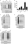
Figure 1.. Protein CoAlation is induced by oxidative stress in mammalian cells and the extent of modification is determined by cellular levels of CoA.
(A) Top, structure of CoA. Bottom, western blot demonstrating specificity of anti-CoA antibody. CoA, dpCoA and 4PP were conjugated to BSA through disulphide linkage and analysed by anti-CoA immunoblot in the presence and absence of DTT. (B) Anti-CoA western blot reveals extensive modification of cellular proteins by CoA in rat cardiomyocytes in response to various oxidising agents. (C) Adult rat cardiomyocytes were untreated or treated with the indicated concentrations of H2O2 for 30 min, and protein CoAlation was examined by anti-CoA immunoblot. (D) The patterns of proteins modified by CoAlation and glutathionylation in response to H2O2 are distinct. Isolated rat hearts were perfused in the presence or absence of 100 µM H2O2 for 20 min, and protein CoAlation and glutathionylation were examined by immunoblotting with anti-CoA or anti-GSH monoclonal antibodies. (E) Comparison of CoA levels in rat tissues, primary and established cell lines. Data are mean ± SEM of at least three biological replicates (n = 5 for cardiomyoctes and HEK293; n = 4 for HepG2 (hepatocellular carcinoma) and MEF (mouse embryonic fibroblasts); n = 3 for the rest). (F) Protein CoAlation is induced by H2O2 in HEK293/Pank1β, but not in parental HEK293 cells. (G) HEK293/Pank1β cells were incubated with the indicated concentrations of H2O2 for 10 min, and protein CoAlation was examined by anti-CoA immunoblot. (H) HEK293/Pank1β cells were incubated with 250 µM H2O2 for the indicated times, and protein CoAlation was examined by anti-CoA immunoblot.
Figure 2.
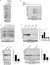
Figure 2.. Protein CoAlation is a reversible post-translational modification and it is prevented by antioxidants.
(A) Anti-CoA western blot showing extensive protein CoAlation in HEK293/Pank1β in response to various oxidising agents. (B) Quantitation of protein-bound CoA after the treatment of HEK293/Pank1β cells with oxidising agents (related to Figure 2A). Values and error bars represent mean ± SEM of at least three independent experiments (n = 6 for control, PAO and diamide; n = 3 for the rest). *P < 0.001 compared with the control group by one-way ANOVA followed by Dunnett's post hoc analysis to correct for multiple comparisons. (C and D) Diamide- and H2O2-induced protein CoAlation in HEK293/Pank1β cells is reversed upon removal of the oxidants. HEK293/Pank1β cells were treated with 250 µM H2O2 and 0.5 mM diamide for 30 min. The medium was then replaced with fresh media without the oxidants and cells were incubated for the indicated times. Protein CoAlation was examined by anti-CoA immunoblot. (E) Protein CoAlation is prevented by antioxidants. HEK293/Pank1β cells were pretreated for 2 h with NAC or Vitamin C before treatment with 0.5 mM diamide for 30 min. Protein CoAlation was examined by anti-CoA western blotting.
Figure 3.
Figure 3.. Induction of protein CoAlation by metabolic stress.
(A) HEK293/Pank1β cells were incubated in pyruvate (pyr) and glucose (glu)-free DMEM medium alone or supplemented with 5 or 20 mM pyruvate. Protein CoAlation was analysed by anti-CoA immunoblot. (B) HEK293/Pank1β cells were incubated in complete pyruvate and glucose-free DMEM alone or supplemented with different concentrations of glucose. Protein CoAlation was analysed by anti-CoA immunoblot. (C) HEK293/Pank1β cells were incubated in pyruvate and glucose-free DMEM alone or supplemented with different concentrations of glucose (glu), galactose (galac), lactate (lac) or acetate (acet). Protein CoAlation was analysed by anti-CoA immunoblot. (D) Induction of protein CoAlation in rat liver after fasting for 24 h. (E) Feeding HF/HS diet for 1 week reduces protein CoAlation in rat liver. (F) Protein CoAlation is decreased in the livers of ob/ob mice. Data are mean ± SEM of three (D and E) or six (F) biological replicates. *P < 0.05 by one-way ANOVA.
Figure 4.
Figure 4.. Proteomic identification of CoAlated proteins in H2O2-treated heart and in the liver of starved rats.
(A) LC–MS/MS spectrum of a CoAlated peptide derived from heart protein, showing an increase in 765 Da, corresponding to covalently bound CoA. The inset (a) shows the structure of CoA and indicates the neutral loss of the ATP moiety (m/z 507 and 427), which can occur under CID (collision-induced dissociation) fragmentation of the precursor ion. The major ions identifying these neutral losses are annotated on the spectrum. (B) Nudix7 cleaves the diphosphate bond of CoA, generating a unique signature for MS/MS analysis. (C and D) Examples of the MS/MS spectra of CoA-modified peptides, corresponding to mitochondrial S-type CK 2 (CKMT2) and GAPDH. Proteins identified to be CoAlated in H2O2-treated heart (E) and liver mitochondria of a 24 h-starved rat (F) were grouped into major functional categories. See Supplementary Tables S1 and S2 for full lists of identified proteins. (G) Major metabolic enzymes are CoAlated in H2O2-treated heart (blue circles) and liver mitochondria of a 24 h-starved rat (red circles), ACC, acetyl CoA carboxylase; MCD, malonyl CoA decarboxylase; CPT1, carnitine palmitoyl transferase 1; CACT, carnitine/acylcarnitine carrier protein; DH, dehydrogenase; CS, citrate synthase; IDH, isocitrate dehydrogenase; OGDH, oxoglutarate dehydrogenase; SDH, succinyl-CoA dehydrogenase; MDH, malate dehydrogenase; PDK, pyruvate dehydrogenase kinase; PDH, pyruvate dehydrogenase; GAPDH, glyceraldehyde 3-phosphate dehydrogenase; SCOT, succinyl-CoA: 3-ketoacid CoA transferase; ACAT1, acetyl-CoA acetyltransferase; I-IV, electron transport chain complexes I–IV; Q, coenzyme Q; CytC; cytochrome C; F0/F1, ATP synthase; ANT, adenine nucleotide translocase; CK, creatine kinase; BDH1, d-β-hydroxybutyrate dehydrogenase; HMGCL, hydroxymethylglutaryl-CoA lyase; HMGCS2, hydroxymethylglutaryl-CoA synthase; ALDH4A1, Δ-1-pyrroline-5-carboxylate dehydrogenase; ALDH5A1, succinate-semialdehyde dehydrogenase; ALDH6A1, methylmalonate-semialdehyde dehydrogenase [acylating]; ALDH7A1, α-aminoadipic semialdehyde dehydrogenase; Glud1, glutamate dehydrogenase 1; Got2, aspartate aminotransferase; GSTZ1, maleylacetoacetate isomerise; HIBADH, 3-hydroxyisobutyrate dehydrogenase; Kat3, kynurenine–oxoglutarate transaminase 3; PCCA, propionyl-CoA carboxylase. Larger-format versions of panels A-G are available in the Supplementary Material.
Figure 5.
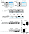
Figure 5.. In vitro and in vivo CoAlation of metabolic enzymes.
(A) In vitro CoAlation of a panel of metabolic enzymes. Recombinant preparations of aconitase, IDH2, PDK2, HMGCS2, as well as CK and GADPH purified from skeletal muscle were incubated with 2–10 mM CoA dimer (CoASSCoA). NEM (25 mM) was added and samples were heated in loading buffer with or without DTT. CoAlation of enzymes was examined by anti-CoA immunoblot. (B) FLAG-tagged CK muscle isoform, GAPDH, IDH2 and PDK2 were transiently overexpressed in HEK293/Pank1β. Transfected cells were treated for 30 min with 0.5 mM H2O2 or 0.5 mM diamide. Overexpressed proteins were immunoprecipitated with an anti-FLAG antibody and immune complexes immunoblotted with anti-CoA antibody. (C and D) Endogenous ACAA2 and HMGCS2 were immunoprecipitated from the liver of control, 24 h starved rats or rats that were fed HF/HS diet for 1 week and analysed by anti-CoA Western blotting. Data are mean ± SEM of three biological replicates. *P < 0.05.
Figure 6.
Figure 6.. Catalytic activities of CK, GAPDH, IDH and PDK2 are modulated by CoAlation.
(A–D) CK, GAPDH purified from skeletal muscle and NADP-dependent IDH purified from heart were incubated with CoASSCoA or GSH dimer (GSSG) and their enzymatic activities assayed spectrophotometrically. The activity of recombinant PDK2 purified from HEK293 cells was assayed radiometrically using PDH as the substrate. Data shown are mean ± SEM of 3 (A, C and D) or 5 (B) independent measurements. Differences between groups were evaluated by a two-way repeated measures ANOVA matching both factors followed by a Tukey post hoc test to correct for multiple comparisons when assessing simple effects or Sidak test when assessing the ‘reducing agent’ effect. *P < 0.05. ns, not significant. (E) Cellular functions of CoA. In addition to its well-established role as an essential metabolic cofactor, CoA may also function as an antioxidant in cellular response to oxidative or metabolic stress via protein CoAlation.

Comment in

Similar articles

Cited by

References

    1. Leonardi R., Zhang Y.-M., Rock C.O. and Jackowski S. (2005) Coenzyme A: back in action. Prog. Lipid Res. 44, 125–153 doi:10.1016/j.plipres.2005.04.001 - DOI - PubMed
    1. Davaapil H., Tsuchiya Y. and Gout I. (2014) Signalling functions of coenzyme A and its derivatives in mammalian cells. Biochem. Soc. Trans. 42, 1056–1062 doi:10.1042/BST20140146 - DOI - PubMed
    1. Srinivasan B. and Sibon O.C.M. (2014) Coenzyme A, more than ‘just’ a metabolic cofactor. Biochem. Soc. Trans. 42, 1075–1079 doi:10.1042/BST20140125 - DOI - PubMed
    1. Theodoulou F.L., Sibon O.C.M., Jackowski S. and Gout I. (2014) Coenzyme A and its derivatives: renaissance of a textbook classic. Biochem. Soc. Trans. 42, 1025–1032 doi:10.1042/BST20140176 - DOI - PubMed
    1. Robishaw J.D., Berkich D. and Neely J.R. (1982) Rate-limiting step and control of coenzyme A synthesis in cardiac muscle. J. Biol. Chem. 257, 10967–10972 PMID: - PubMed

Publication types

LinkOut - more resources