. 2017 Mar 25:23:1448-1455.
doi: 10.12659/msm.899804.

Berberine Restricts Coxsackievirus B Type 3 Replication via Inhibition of c-Jun N-Terminal Kinase (JNK) and p38 MAPK Activation In Vitro

Affiliations

Berberine Restricts Coxsackievirus B Type 3 Replication via Inhibition of c-Jun N-Terminal Kinase (JNK) and p38 MAPK Activation In Vitro

Qian Dai et al. Med Sci Monit. .

Abstract

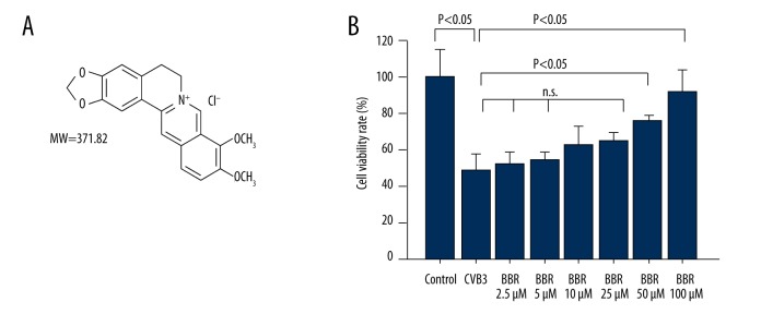
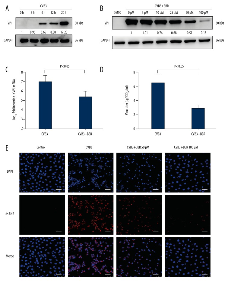
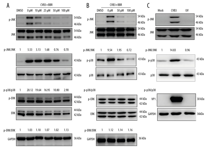
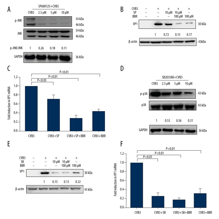
BACKGROUND At present, the treatment of coxsackievirus-induced myocarditis remains difficult. Berberine (BBR), an isoquinoline alkaloid isolated from traditional medicine herbs, exhibits significant anti-viral efficacy against various viruses. However, the underlying mechanism by which BBR controls CVB3 infection has not yet been reported. The purpose of this study was to investigate the anti-viral efficacy of BBR against CVB3 infection and its mechanism. MATERIAL AND METHODS In our experiments, the protein levels of VP1 and MAPKs signal pathway were measured by Western blot. The mRNA level of VP1 was measured by RT-PCR. The virus titers were determined by TCID50 assay. RESULTS We found that BBR treatment significantly decreased CVB3 replication in HeLa cells. In addition, the BBR treatment reduced the phosphorylation levels of JNK and p38 MAPK upon CVB3 infection in both HeLa cells and primary rat myocardial cells. CONCLUSIONS Taken together, these results suggest that BBR inhibits CVB3 replication through the suppression of JNK and p38 MAPK activation, shedding new light on the investigation of therapeutic strategies against CVB3-induced viral myocarditis.


Figures

Figure 1
Figure 1
Effects of BBR on HeLa cell viability. (A) The chemical structure of BBR. (B) The cell viability in CVB3-infected or BBR-treated cells were determined by CCK-8 assay. Data show mean ±SD from 3 independent experiments. n.s. indicates no significance.
Figure 2
Figure 2
BBR inhibits CVB3 replication in vitro. (A) HeLa cells were infected with CVB3 at 3 MOI for 1 h. Cell lysates were collected at the indicated times (3 h, 6 h, 12 h, 20 h) post-infection. Western blot analysis was performed to detect the expression of virus VP1 protein. (B) At 1 h after HeLa cells were infected with CVB3 (MOI=3), they were treated with BBR at the indicated concentrations (3 μM, 10 μM, 25 μM, 50 μM, 100 μM) for 20 h. Cell lysates were harvested and subjected to further analyses. Western blot analysis was performed to evaluate the expression of virus VP1 protein. (C) At 1 h after HeLa cells were infected with CVB3 (MOI=3), they were treated with BBR (100 μM) for 20 h. Real-time PCR analysis was performed to evaluate the levels of virus vp1 mRNA. (D) The viral titers after BBR (100 μM) treatment were determined by the Reed-Muench method. (E) IF staining was performed to detect the production of virus dsRNA during virus replication (dsRNA, red; nuclei, blue). The scale bar is 25 μm. Representative data from triplicate experiments are shown. Data show mean ±SD from 3 independent experiments. GAPDH was used as a protein loading control or endogenous reference gene.
Figure 3
Figure 3
BBR inhibited CVB3-induced activation of JNK and p38. HeLa cells and primary myocardial cells infected with CVB3 (MOI=3) for 1 h were treated with BBR at the indicated concentrations (10 μM, 25 μM, 50 μM, 100 μM) for 20 h. The phosphorylation levels of JNK, p38, and ERK in HeLa cells (A) and in primary myocardial cells (B) were analyzed by Western blot. The controls were treated with vehicle control (DMSO, 0.1%). (C) HeLa cells were infected with mock-control, CVB3, or UV-inactivated CVB3 (MOI=3). Representative data from triplicate experiments are shown. GAPDH was used as a loading control.
Figure 4
Figure 4
The inhibition of JNK or p38 activation by its corresponding inhibitor or BBR reduces CVB3 replication. HeLa cells infected with CVB3 (MOI=3) for 1 h were treated with JNK inhibitor SP600125 (SP, 2.5 μM, 5 μM, or 10 μM) or p38 inhibitor SB203580 (SB, 2.5 μM, 5 μM, or 10 μM) for 20 h. The phosphorylation levels of JNK or p38 were analyzed by Western blot (A, D). HeLa cells were infected with either mock-control or CVB3 (MOI=3). At 1 h after infection, the virus-infected cells treated with SP or SB (10 μM) were concurrently treated with or without BBR (100 μM) for 20 h. The expression of CVB3 VP1 was determined by Western blot (B, E) and real-time PCR (C, F). Representative data from triplicate experiments are shown in A, B, D, and E. GAPDH and β-actin were used as the loading control. Data in C and F show mean ±SD from 3 independent experiments.

References

    1. Aretz HT, Billingham ME, Edwards WD, et al. Myocarditis. A histopathologic definition and classification. Am J Cardiovasc Pathol. 1987;1:3–14. - PubMed
    1. Esfandiarei M, McManus BM. Molecular biology and pathogenesis of viral myocarditis. Ann Rev Pathol. 2008;3:127–55. - PubMed
    1. Grist NR, Reid D. Organisms in myocarditis/endocarditis viruses. J Infect. 1997;34:155. - PubMed
    1. Baboonian C, Davies MJ, Booth JC, McKenna WJ. Coxsackie B viruses and human heart disease. Curr Top Microbiol Immunol. 1997;223:31–52. - PubMed
    1. Okada I, Matsumori A, Matoba Y, et al. Combination treatment with ribavirin and interferon for coxsackievirus B3 replication. J Lab Clin Med. 1992;120:569–73. - PubMed

MeSH terms