

Posterior cingulate cross-hemispheric functional connectivity predicts the level of consciousness in traumatic brain injury
- PMID: 28341824
- PMCID: PMC5428308
- DOI: 10.1038/s41598-017-00392-5
Posterior cingulate cross-hemispheric functional connectivity predicts the level of consciousness in traumatic brain injury
Abstract
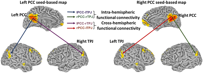
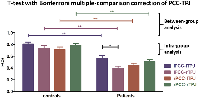
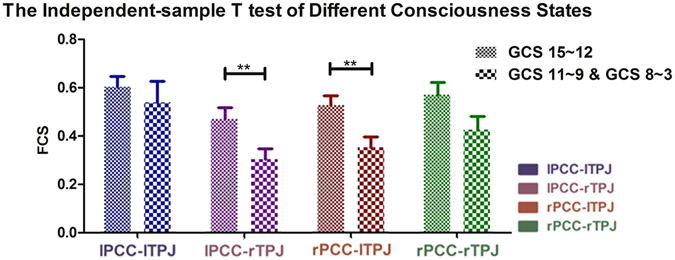
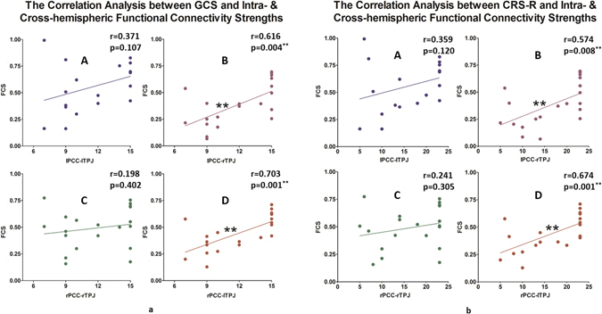
Previous studies have demonstrated that altered states of consciousness are related to changes in resting state activity in the default-mode network (DMN). Anatomically, the DMN can be divided into anterior and posterior regions. The anterior DMN includes the perigenual anterior cingulate cortex and other medial prefrontal cortical regions, whereas the posterior DMN includes regions such as the posterior cingulate cortex (PCC) and the temporal parietal junction (TPJ). Although differential roles have been attributed to the anterior and posterior DMN regions, their exact contributions to consciousness levels remain unclear. To investigate the specific role of the posterior DMN in consciousness levels, we investigated 20 healthy controls (7 females, mean age = 33.6 years old) and 20 traumatic brain injury (TBI) patients (5 females, mean age = 43 years old) whose brain lesions were mainly restricted to the bilateral frontal cortex but retained a well-preserved posterior DMN (e.g., the PCC and the TPJ) and who exhibited varying levels of consciousness. We investigated the intra- and cross-functional connectivity strengths (FCSs) between the right/left PCC and the right/left TPJ and their correlation with consciousness levels. Significant reductions in both the intra- and cross-hemispheric FCSs were observed in patients compared with controls. A significant correlation with consciousness levels was observed only for the cross-hemispheric PCC-TPJ FCS but not for the intra-hemispheric PCC-TPJ FCS. Taken together, our results show that the cross-hemispheric posterior DMN is related to consciousness levels in a specific group of patients without posterior structural lesions. We therefore propose that the PCC may be central in maintaining consciousness through its cross-hemispheric FC with the TPJ.
Conflict of interest statement
The authors declare that they have no competing interests.
Figures
Similar articles
-
[Altered patterns of functional connectivity of posterior cingulate cortex on resting-state magnetic resonance imaging in children with attention-deficit or hyperactivity disorder].Zhonghua Yi Xue Za Zhi. 2013 Jun 25;93(24):1881-5. Zhonghua Yi Xue Za Zhi. 2013. PMID: 24124739 Chinese.
-
Intrinsic Functional Connectivity Patterns Predict Consciousness Level and Recovery Outcome in Acquired Brain Injury.J Neurosci. 2015 Sep 16;35(37):12932-46. doi: 10.1523/JNEUROSCI.0415-15.2015. J Neurosci. 2015. PMID: 26377477 Free PMC article.
-
The structural and functional connectivity of the posterior cingulate cortex: comparison between deterministic and probabilistic tractography for the investigation of structure-function relationships.Neuroimage. 2014 Nov 15;102 Pt 1:118-27. doi: 10.1016/j.neuroimage.2013.12.022. Epub 2013 Dec 21. Neuroimage. 2014. PMID: 24365673 Review.
-
Alteration of the Intra- and Cross- Hemisphere Posterior Default Mode Network in Frontal Lobe Glioma Patients.Sci Rep. 2016 Jun 1;6:26972. doi: 10.1038/srep26972. Sci Rep. 2016. PMID: 27248706 Free PMC article.
-
Application of Functional MRI in Parkinson's Disease and Default Mode Network: Review of the Literature.Can J Neurol Sci. 2025 May 19:1-8. doi: 10.1017/cjn.2025.110. Online ahead of print. Can J Neurol Sci. 2025. PMID: 40384213 Review.
Cited by
-
Alteration in ventral tegmental area and default mode network interplay and prediction of coma recovery in patients with sTBI.Heliyon. 2023 Apr 5;9(4):e15279. doi: 10.1016/j.heliyon.2023.e15279. eCollection 2023 Apr. Heliyon. 2023. PMID: 37128308 Free PMC article.
-
Fast and Slow Recovery of Consciousness Following Traumatic Brain Injury.Neurocrit Care. 2025 Jun 27. doi: 10.1007/s12028-025-02304-2. Online ahead of print. Neurocrit Care. 2025. PMID: 40579677
-
Dissociation of the subjective and objective bodies: Out-of-body experiences following the development of a posterior cingulate lesion.J Neuropsychol. 2020 Mar;14(1):183-192. doi: 10.1111/jnp.12199. Epub 2019 Dec 21. J Neuropsychol. 2020. PMID: 31863565 Free PMC article.
-
Enhanced brain parcellation via abnormality inpainting for neuroimage-based consciousness evaluation of hydrocephalus patients by lumbar drainage.Brain Inform. 2023 Jan 19;10(1):3. doi: 10.1186/s40708-022-00181-5. Brain Inform. 2023. PMID: 36656455 Free PMC article.
-
Abnormalities of cortical and subcortical spontaneous brain activity unveil mechanisms of disorders of consciousness and prognosis in patients with severe traumatic brain injury.Int J Clin Health Psychol. 2024 Oct-Dec;24(4):100528. doi: 10.1016/j.ijchp.2024.100528. Epub 2024 Nov 28. Int J Clin Health Psychol. 2024. PMID: 39659957 Free PMC article.
References
-
- Uddin LQ, Supekar KS, Ryali S, Menon V. Dynamic reconfiguration of structural and functional connectivity across core neurocognitive brain networks with development. The Journal of neuroscience: the official journal of the Society for Neuroscience. 2011;31:18578–18589. doi: 10.1523/JNEUROSCI.4465-11.2011. - DOI - PMC - PubMed
Publication types
MeSH terms
Grants and funding
LinkOut - more resources
Full Text Sources
Other Literature Sources
Medical
