Observational Study
. 2017 Mar 24;7(1):414.
doi: 10.1038/s41598-017-00345-y.

The relationship between retinal nerve fibre layer thickness profiles and CorvisST tonometry measured biomechanical properties in young healthy subjects

Affiliations
Observational Study

The relationship between retinal nerve fibre layer thickness profiles and CorvisST tonometry measured biomechanical properties in young healthy subjects

Masato Matsuura et al. Sci Rep. .

Abstract

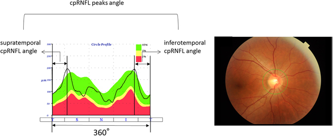
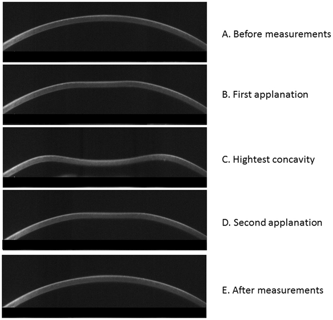
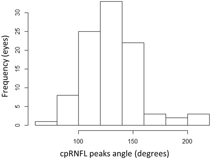
We previously reported that a shallow circumpapillary retinal nerve fiber layer (cpRNFL) peak angle as measured by optical coherence tomography (OCT) suggests the temporal retina is stretched around the optic disc from the papillo-macular bundle (Yamashita T et al. Investigative Ophthalmol Vis Sci 2013). The purpose of the current study was to investigate the relationship between CorvisST tonometry (CST) corneal measurements, axial length (AL) and the change in OCT-measured cpRNFL peak angle, in young healthy subjects. OCT and CST measurements were carried out in 97 eyes of 97 young healthy volunteers. The relationship between cpRNFL peak angle and 12 CST parameters, adjusted for AL, was investigated using linear modelling. The mean ± standard deviation cpRNFL peak angle of the 97 healthy volunteers was 130.6 ± 25.4 (range: 77.8 to 207.0) degrees. The optimal linear model to explain cpRNFL peak angle (chosen from 216 different models) included three CST variables related to the speed and size of energy absorption (namely, A1 time, A1 length and A2 time), in addition to AL. In eyes with longer AL and shorter energy absorption in CST measurement, temporal retina is stretched around the optic disc from the papillo-macular bundle, as suggested by a shallow cpRNFL peak angle.


Conflict of interest statement

The authors declare that they have no competing interests.

Figures

Figure 1
Figure 1
Calculation of cpRNFL peaks angle. The cpRNFL peaks angle was calculated as the angle between the supratemporal cpRNFL peak angle and inferotemporal cpRFNL peak angle. cpRNFL: circumpapillary retinal nerve fiber layer.
Figure 2
Figure 2
Corneal movement during the CST measurement. In the CST tonometry measurement, a rapid air puff is applied to cornea and cornea moves inward. The figures show the corneal shape in each phase: (A) Prior to air puff applanation, (B) first applanation, (C) highest concavity, (D) second applanation, and (E) posterior to air puff applanation. CST: Corvis ST tonometry.
Figure 3
Figure 3
Histogram of cpRNFL peaks angle. The cpRNFL peaks angle was 130.6 ± 25.4 (mean ± standard deviation) [range: 77.8 to 207.0] degrees. cpRNFL: circumpapillary retinal nerve fiber layer.

References

    1. Quigley HA. Number of people with glaucoma worldwide. Br J Ophthalmol. 1996;80:389–393. doi: 10.1136/bjo.80.5.389. - DOI - PMC - PubMed
    1. Congdon N, et al. Causes and prevalence of visual impairment among adults in the United States. Arch Ophthalmol. 2004;122:477–485. doi: 10.1001/archopht.122.4.477. - DOI - PubMed
    1. Mistlberger A, et al. Heidelberg retina tomography and optical coherence tomography in normal, ocular-hypertensive, and glaucomatous eyes. Ophthalmology. 1999;106:2027–2032. doi: 10.1016/S0161-6420(99)90419-0. - DOI - PubMed
    1. Zangwill LM, Williams J, Berry CC, Knauer S, Weinreb RN. A comparison of optical coherence tomography and retinal nerve fiber layer photography for detection of nerve fiber layer damage in glaucoma. Ophthalmology. 2000;107:1309–1315. doi: 10.1016/S0161-6420(00)00168-8. - DOI - PubMed
    1. Leung CK, et al. Retinal nerve fiber layer measurements in myopia: An optical coherence tomography study. Invest Ophthalmol Vis Sci. 2006;47:5171–5176. doi: 10.1167/iovs.06-0545. - DOI - PubMed

Publication types

LinkOut - more resources