. 2017 Mar 24;7(1):396.
doi: 10.1038/s41598-017-00313-6.

FAM46C is critical for the anti-proliferation and pro-apoptotic effects of norcantharidin in hepatocellular carcinoma cells

Affiliations

FAM46C is critical for the anti-proliferation and pro-apoptotic effects of norcantharidin in hepatocellular carcinoma cells

Qiao-Yan Zhang et al. Sci Rep. .

Erratum in

Abstract

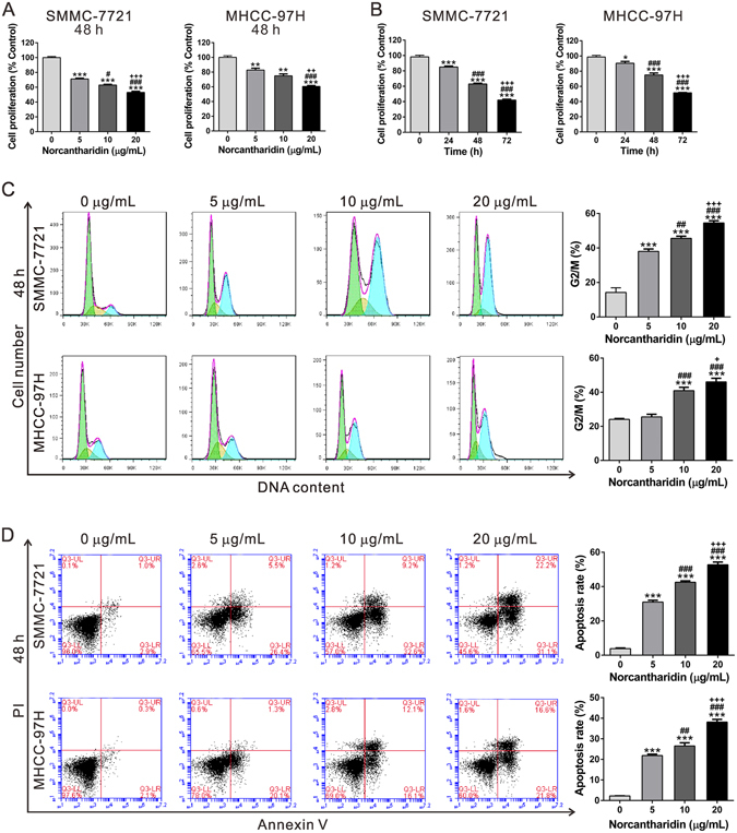
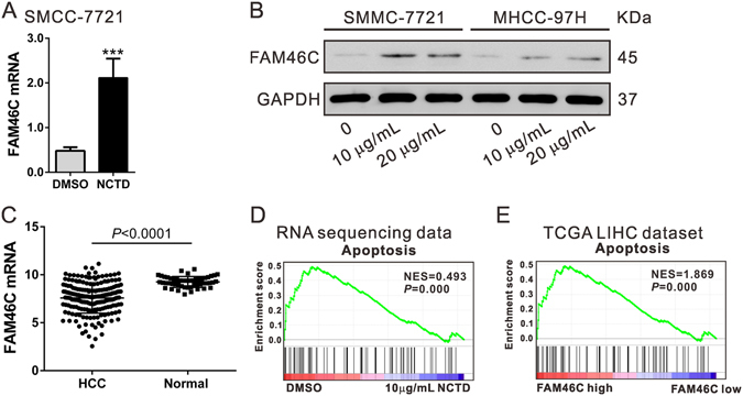
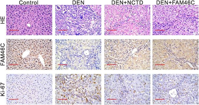
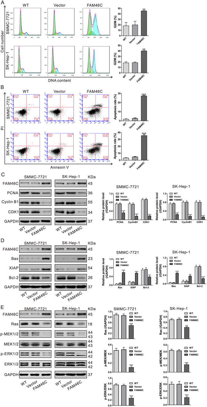
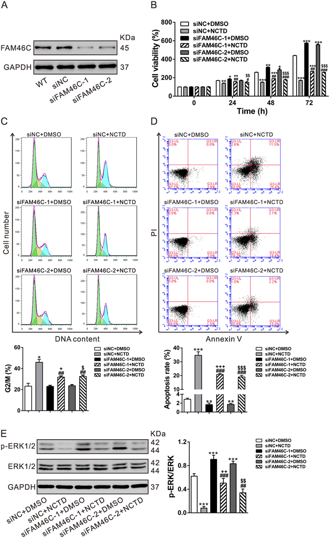
Norcantharidin (NCTD), a demethylated analog of cantharidin derived from Chinese traditional medicine blister beetle, has been currently used as an anticancer drug for various cancers including hepatocellular carcinoma (HCC). In this study, for a more comprehensive understanding of the targets of NCTD in HCC, next-generation RNA-Seq was utilized. We revealed that the expression of FAM46C, which has been reported as a tumor suppressor for multiple myeloma, was enhanced after NCTD treatment. Re-analysis of TCGA (The Cancer Genome Atlas) LIHC (liver hepatocellular carcinoma) dataset demonstrated that FAM46C expression was significantly lower in HCC tissues than in normal liver tissues. NCTD injection or FAM46C overexpression could mitigate diethylnitrosamine (DEN)-initiated HCC in mice. Ectopic expression of FAM46C in two HCC cell lines, SMCC-7721 and SK-Hep-1, significantly repressed cell proliferation, and increased cells population in G2/M phase and cell apoptotic rate. We also found that FAM46C overexpression caused a notable decrease in Ras expression, MEK1/2 phosphorylation and ERK1/2 phosphorylation. More importantly, FAM46C knockdown significantly weakened the biological effects of NCTD on HCC cells, which suggested NCTD exerted the anticancer functions partially through up-regulating FAM46C. In conclusion, FAM46C, a tumor suppressor for HCC, is important for the anti-proliferation and proapoptotic effects of NCTD.


Conflict of interest statement

The authors declare that they have no competing interests.

Figures

Figure 1
Figure 1
Effects of NCTD on cell proliferation and apoptosis of SMCC-7721 and MHCC-97H cells. (A) SMCC-7721 and MHCC-97H cells were treated with DMSO or NCTD (5, 10 and 20 µg/mL) for 48 h. CCK-8 assay was carried out to assess cell proliferation. The relative cell proliferation was defined as the percentage of cells treated with DMSO (% Control). **P < 0.01, ***P < 0.001 as compared with DMSO group; # P < 0.05, ### P < 0.001 as compared with 5 µg/mL NCTD-treated group; ++ P < 0.01, +++ P < 0.001 as compared with 10 µg/mL NCTD-treated group. (B) The cells were treated by 10 μg/mL NCTD for 24, 48 and 72 h. At the end of incubation, CCK-8 assay was carried out to assess cell proliferation. The relative cell proliferation was expressed as the percentage of OD450 compared with that of the control (% Control). *P < 0.05, ***P < 0.001 as compared with 0 h; ### P < 0.001 as compared with 24 h; +++ P < 0.001 as compared with 48 h. (C,D) SMCC-7721 and MHCC-97H cells were treated with DMSO or NCTD for 48 h. Cell cycle (C) distribution was assessed by PI staining and flow cytometric analysis. Cell percentages in G2/M phase were shown here. Cell apoptosis (D) was evaluated by Annexin V-FITC/PI staining followed by flow cytometric analysis. Cells in the lower right quadrant are Annexin V-positive and PI-negative staining, representing the early apoptotic cells. ***P < 0.001 as compared with DMSO group; ## P < 0.01, ### P < 0.001 as compared with 5 µg/mL NCTD-treated group; +++ P < 0.001 as compared with 10 µg/mL NCTD-treated group. All experiments shown were performed independently at least three times.
Figure 2
Figure 2
Effects of NCTD on FAM46C expression. (A) RNA-sequencing data showed that NCTD significantly induced FAM46C mRNA expression. (B) SMCC-7721 and MHCC-97H cells were treated with DMSO or NCTD (10 and 20 µg/mL) for 48 h. The protein level of FAM46C was significantly induced by NCTD treatment. (C) FAM46C was down-regulated in HCC tissues (n = 191) compared to the normal tissues (n = 50) in TCGA LIHC dataset. (D) GSEA revealed that the cell apoptosis pathway was closely related with NCTD treatment. Black bars indicated individual genes in rank order. NES, normalized enrichment score. (E) GSEA was performed using TCGA LIHC dataset. The cell apoptosis pathway was strongly associated with FAM46C expression.
Figure 3
Figure 3
Pathological changes of liver in 4 groups mice (n = 8 per group). Light microscopy of HE staining, and IHC staining of FAM46C and Ki-67 (200x magnification) in Control group, DEN group, DEN+NCTD group and DEN+FAM46C group.
Figure 4
Figure 4
FAM46C overexpression inhibited HCC cell proliferation. (A) Expression of FAM46C in 6 HCC cell lines as determined by Western blotting. (B) Ectopic expression of FAM46C in SMCC-7721 and SK-Hep-1 cells was examined by Western blotting. (C) Ectopic expression of FAM46C significantly decreased cell proliferation as determined by CCK-8 assay. All experiments shown were performed independently at least three times. ***P < 0.001 as compared with Vector virus-infected cells.
Figure 5
Figure 5
FAM46C overexpression resulted in G2/M phase arrest (A) and a significant increase of cell apoptosis (B) as determined by flow cytometry analyses at 48 h after viral infection. ***P < 0.001 as compared with Vector virus-infected cells. (C) Protein levels of FAM46C and cell growth-related factors (PCNA, Cyclin B1 and CDK1) were evaluated by Western blotting. (D) Protein levels of FAM46C and apoptotic factors (Bcl-2, Bax and XIAP) were assessed. (E) The levels of Ras, p-MEK1/2, MEK, p-ERK1/2 and ERK1/2 were evaluated. All experiments shown were performed independently at least three times. **P < 0.01 and ***P < 0.001 as compared with Vector virus-infected cells.
Figure 6
Figure 6
FAM46C is critical for the anti-cell growth effects of NCTD on HCC cells. (A) MHCC-97H cells were transfected with siNC, siFAM46C-1 or siFAM46C-2. At 48 h after transfection, FAM46C protein expression was detected by Western blotting. (B,C,D) MHCC-97H cells were divided into 6 groups: siNC+DMSO, siNC+NCTD (10 μg/mL), siFAM46C-1+DMSO, siFAM46C-1+NCTD (10 μg/mL), siFAM46C-2+DMSO and siFAM46C-2+NCTD (10 μg/mL). Cell proliferation (B) was evaluated by CCK-8 assay at indicated time points. At 48 h after treatment, cell percentages in G2/M phase (C) and cell apoptosis (D) were detected by flow cytometric analysis. (E) Western blot analysis of ERK phosphorylation at 48 h after treatment. All experiments shown were performed independently at least three times. *P < 0.05, **P < 0.01, ***P < 0.001 as compared with siNC+DMSO; ## P < 0.01, ### P < 0.001 as compared with siNC+NCTD; + P < 0.001, ++ P < 0.05,+++ P < 0.001 as compared with siFAM46C-1+NCTD; $ P < 0.001, $$ P < 0.05, $$$ P < 0.001 as compared with siFAM46C-2+NCTD.

References

    1. El-Serag HB, Marrero JA, Rudolph L, Reddy KR. Diagnosis and treatment of hepatocellular carcinoma. Gastroenterology. 2008;134:1752–1763. doi: 10.1053/j.gastro.2008.02.090. - DOI - PubMed
    1. Torre LA, et al. Global cancer statistics, 2012. CA Cancer J Clin. 2015;65:87–108. doi: 10.3322/caac.21262. - DOI - PubMed
    1. Poon RT. Optimal initial treatment for early hepatocellular carcinoma in patients with preserved liver function: transplantation or resection? Ann Surg Oncol. 2007;14:541–547. doi: 10.1245/s10434-006-9156-z. - DOI - PubMed
    1. Bruix J, Sherman M. Management of hepatocellular carcinoma: an update. Hepatology. 2011;53:1020–1022. doi: 10.1002/hep.24199. - DOI - PMC - PubMed
    1. Wang GS. Medical uses of mylabris in ancient China and recent studies. J Ethnopharmacol. 1989;26:147–162. doi: 10.1016/0378-8741(89)90062-7. - DOI - PubMed

Publication types

MeSH terms