. 2017 Mar 24;7(1):389.
doi: 10.1038/s41598-017-00334-1.

Embryonic defence mechanisms against glucose-dependent oxidative stress require enhanced expression of Alx3 to prevent malformations during diabetic pregnancy

Affiliations

Embryonic defence mechanisms against glucose-dependent oxidative stress require enhanced expression of Alx3 to prevent malformations during diabetic pregnancy

Patricia García-Sanz et al. Sci Rep. .

Abstract

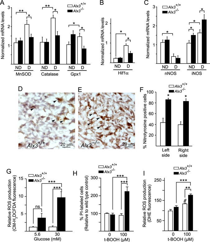
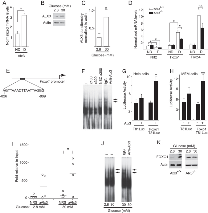
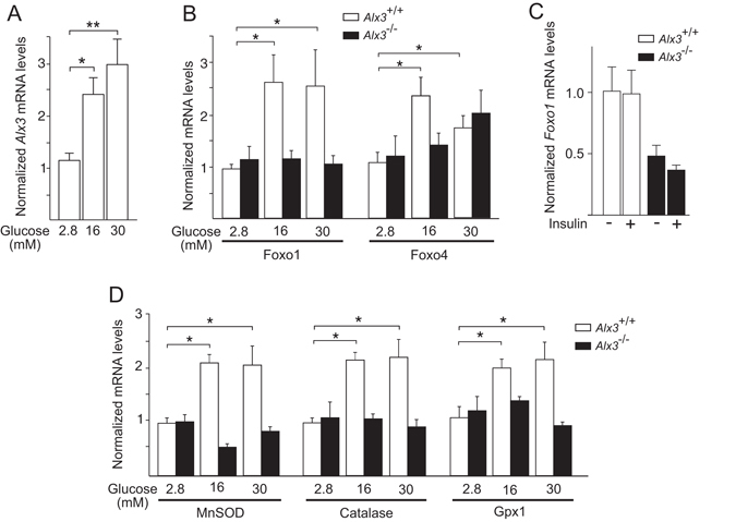
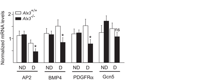
Oxidative stress constitutes a major cause for increased risk of congenital malformations associated to severe hyperglycaemia during pregnancy. Mutations in the gene encoding the transcription factor ALX3 cause congenital craniofacial and neural tube defects. Since oxidative stress and lack of ALX3 favour excessive embryonic apoptosis, we investigated whether ALX3-deficiency further increases the risk of embryonic damage during gestational hyperglycaemia in mice. We found that congenital malformations associated to ALX3-deficiency are enhanced in diabetic pregnancies. Increased expression of genes encoding oxidative stress-scavenging enzymes in embryos from diabetic mothers was blunted in the absence of ALX3, leading to increased oxidative stress. Levels of ALX3 increased in response to glucose, but ALX3 did not activate oxidative stress defence genes directly. Instead, ALX3 stimulated the transcription of Foxo1, a master regulator of oxidative stress-scavenging genes, by binding to a newly identified binding site located in the Foxo1 promoter. Our data identify ALX3 as an important component of the defence mechanisms against the occurrence of developmental malformations during diabetic gestations, stimulating the expression of oxidative stress-scavenging genes in a glucose-dependent manner via Foxo1 activation. Thus, ALX3 deficiency provides a novel molecular mechanism for developmental defects arising from maternal hyperglycaemia.


Conflict of interest statement

The authors declare that they have no competing interests.

Figures

Figure 1
Figure 1
Induction of diabetic pregnancies in wild type and Alx3-deficient mice. (A) Basal blood glucose concentrations after fasting in wild-type (n = 27) and Alx3-deficient (n = 38) female mice. **P < 0.01, Student’s t-test. (B) Glucose tolerance tests carried out in fasting wild-type (left panel, blue, n = 21) or Alx3-deficient (right panel, red, n = 19) female mice. Both non-pregnant (clear squares) and pregnant mice (solid squares) at 8.5 days post coitus were tested. *P < 0.05, **P < 0.01 relative to non pregnant wild type animals at each time point (ANOVA followed by Bonferroni transformation). No statistically significant differences were found between pregnant and non-pregnant Alx3-null females. (C) Blood glucose concentrations after fasting in wild type (white circles, n = 22) and Alx3-deficient (black circles, n = 43) female mice. Injections of streptozotocin (STZ) and subcutaneous implantation of insulin pellets (Ins) are indicated by arrows. Note that severe hyperglycaemia develops again during pregnancy up to 10.5 days of gestation. (D) Number of embryos per litter observed in non-diabetic (ND) or diabetic (D) pregnancies at 10.5 days of gestation. The numbers of litters were: Alx3 +/+, 20 (ND) and 15 (D); Alx3 −/−, 19 (ND) and 29 (D). Pregnancies without any embryos were not counted for litter size calculations. *p < 0.05; **P < 0.01, Student’s t-test. All values represent mean + s.e.m.
Figure 2
Figure 2
Malformations observed in embryos from diabetic pregnancies after 10.5 days of gestation. (A,B) Examples of caudal malformations observed in wild type embryos from diabetic pregnancies. Tail flexion defects are indicated by arrowheads. (D,E) Examples of cranial malformations observed in heterozygote Alx3-deficient embryos from diabetic pregnancies. These include defects of the rostral mesenchyme affecting midline closure at the level of the forehead (D, arrowheads), defects at the level of the mesencephalic vesicles (D, arrows) and defects of facial mesenchyme expansion (E, arrowheads) affecting the development of the telencephalic vesicles (E, arrows). (G,H) Examples of severe malformations found in Alx3-null embryos developing in diabetic mothers. Malformations affected several embryonic regions including the forehead mesenchyme, the branchial arches and the cranial neural tube (G), or were restricted to severe craniofacial and cranial neural tube closure defects (H, arrows). Arrowheads indicate open neural folds. (C,F and I) depict normal wild type embryos for comparisons. Scale bars represent 1 mm in all cases except in G (1.5 mm). Abbreviations: ba, Branchial arches; FLB, Forelimb bud; HLB, Hind limb bud; m, Mesencephalon; r, Rhombencephalon; t, Telencephalic vesicles.
Figure 3
Figure 3
Impaired activation of genes encoding oxidative stress-scavenging enzymes and increased oxidative stress in Alx3-deficient embryos developing during diabetic pregnancies. (AC) Relative levels of mRNA extracted from embryos of non-diabetic (ND) or diabetic (D) wild type (white bars) or Alx3-deficient (black bars) mice, as assessed by quantitative RT-PCR after 10.5 days of gestation. Shown are mRNAs encoding manganese superoxide dismutase (MnSOD), catalase and glutathione peroxidase-1 (Gpx1) (A), Hif1α (B) or neuronal (n) or inducible (i) nitric oxide synthase (NOS) (C). *P < 0.05; **P < 0.01; Student’s t-test (n = 7 per group). (D and E) Representative sections showing nitrotyrosine immunohistochemistry in the head mesenchyme of Alx3 +/+ (D) or Alx3 −/− (E) embryos from diabetic pregnancies. Arrowheads indicate representative examples of nitrotyrosine-positive cells. (F) Quantification of the percentage of nitrotyrosine-positive cells detected by immunohistochemistry in sections from wild type (white columns) or Alx3-deficient (black columns) embryos from diabetic pregnancies. Equivalent mesenchyme regions symmetrically located at both left and right sides of each section relative to the midline of the embryo were scored independently. Approximately 1000 cells per section from 6 sections obtained from 3 embryos in each group were counted. *P < 0.001, Student’s t-test. (G) Quantitation of ROS in control (white bars) or Alx3-deficient (black bars) primary MEM cells cultured in the presence of the indicated concentrations of glucose and labelled with the fluorescent dye CM-H2DCFDA (n = 6 per group). (H and I) Response of control or Alx3-deficient primary MEM cells to t-BOOH treatment. In H, the relative numbers of non-viable cells identified by propidium iodide (PI) staining is represented (n = 14 for untreated cell groups; n = 12 for t-BOOH-treated cell groups). In I, ROS production as measured by DHE fluorescence is shown (n = 7 for untreated cell groups; n = 6 for t-BOOH-treated cell groups). In (GI), *P < 0.05; **P < 0.01; ***P < 0.001; # P < 0.05, compared with untreated wild type cells. ANOVA followed by Bonferroni test. All values represent mean + s.e.m.
Figure 4
Figure 4
Regulation of the Foxo1 promoter by ALX3. (A) Quantitative RT-PCR of Alx3 mRNA in embryos from non-diabetic (ND) or diabetic (D) wild type mice (n = 7 per group). (B) Western blot showing ALX3 in primary MEM cells cultured in the indicated concentrations of glucose. (C) Densitometric measurements of ALX3 bands relative to actin bands from three western blots similar to that shown in B. D) Quantitative RT-PCR of Nrf2, Foxo1 or Foxo4 mRNAs, extracted from embryos of non-diabetic (ND) or diabetic (D) wild type (white bars) or Alx3-deficient (black bars) mice; n.s., non-significant (n = 7 per group). (E) ALX3-binding site and its position in the mouse Foxo1 promoter. (F) Electrophoretic mobility shift assays showing the binding of proteins from nuclear extracts of primary MEM cells to an oligonucleotide probe containing the sequence indicated in E. The absence (−) or presence of competing or nonspecific competing (NSC) oligonucleotides at the indicated fold molar excess, or of an ALX3 antibody or control IgG, is depicted on top. Arrows indicate complexes containing ALX3. (G,H) Relative luciferase activity elicited in Hela (G) or primary Alx3-defcient MEM (H) cells co-transfected with the reporter plasmids Foxo1T81Luc or control pT81Luc, and an ALX3 expression plasmid (n = 5). (I) Quantitative PCR amplification of Foxo1 chromatin immunoprecipitated with anti-ALX3 antiserum or with control non-immune rabbit serum (NRS) from primary MEM cells isolated from wild type mice and cultured in the presence of the indicated concentrations of glucose. Horizontal lines represent the mean (n = 4). (J) Electrophoretic mobility shift assays showing binding of nuclear extracts from primary MEM cells cultured at the indicated concentrations of glucose to the Foxo1 promoter site indicated in E. Arrows indicate the presence of ALX3. (K) Western blot showing FOXO1 in primary wild type (left panels) or Alx3-deficient (right panel) MEM cells cultured in the indicated concentrations of glucose. A representative example of three independent experiments with similar results is depicted. Uncropped blots are provided in Supplementary Fig. S5. Except in I, all values represent mean + s.e.m. *P < 0.05, **P < 0.01, Student’s t-test.
Figure 5
Figure 5
Impaired response to glucose in Alx3-deficient cells. (A) Relative levels of Alx3 mRNA in primary MEM cells obtained from wild type mice cultured in the presence of the indicated concentrations of glucose (n = 5). (B) Relative levels of Foxo1 and Foxo4 mRNA in primary MEM cells obtained from wild type (white bars) or Alx3-deficient (black bars) embryos cultured in the presence of the indicated glucose concentrations (n = 7 per group). (C) Relative levels of Foxo1 mRNA in primary MEM cells from wild type (white bars) or Alx3-deficient (black bars) embryos cultured in the presence of 0.5% FBS without (−) or with (+) insulin (100 nM) (n = 5 per group). (D) Relative levels of MnSOD, catalase and Gpx1 mRNA in primary MEM cells from wild type (white bars) or Alx3-deficient (black bars) embryos cultured in the indicated glucose concentrations (n = 7 per group). All data obtained by quantitative RT-PCR. In all cases, values represent mean + s.e.m. *p < 0.05, **p < 0.01, Student’s t-test.
Figure 6
Figure 6
Altered gene expression in Alx3-deficient embryos from diabetic pregnancies. Relative levels of mRNA encoding representative developmentally regulated genes in wild type (white bars) or Alx3-null (black bars) embryos obtained from non-diabetic (ND) or diabetic (D) mothers. Values represent mean + s.e.m. *p < 0.05, Student’s t-test (n = 7 per group).

Similar articles

Cited by

References

    1. Farrell T, Neale L, Cundy T. Congenital anomalies in the offspring of women with type1, type 2 and gestational diabetes. Diabet. Med. 2002;19:322–326. doi: 10.1046/j.1464-5491.2002.00700.x. - DOI - PubMed
    1. Tennant PWG, Glinianaia SV, Bilous RW, Rankin J, Bell R. Pre-existing diabetes, maternal glycated haemoglobin, and the risks of fetal and infant death: a population-based study. Diabetologia. 2014;57:285–294. doi: 10.1007/s00125-013-3108-5. - DOI - PubMed
    1. Martínez-Frías ML. Epidemiological analysis of outcomes of pregnancy in diabetic mothers: identification of the most characteristic and most frequent congenital anomalies. Am. J. Med. Genet. 1994;51:108–113. doi: 10.1002/ajmg.1320510206. - DOI - PubMed
    1. Simeone RM, et al. Diabetes and congenital heart defects: a systematic review, meta-analysis, and modeling project. Am J Prev Med. 2015;48:195–204. doi: 10.1016/j.amepre.2014.09.002. - DOI - PMC - PubMed
    1. Fine EL, Horal M, Chang TI, Fortin G, Loeken MR. Evidence that elevated glucose causes altered gene expression, apoptosis, and neural tube defects in a mouse model of diabetic pregnancy. Diabetes. 1999;48:2454–2462. doi: 10.2337/diabetes.48.12.2454. - DOI - PubMed

Publication types

MeSH terms