. 2017 May;133(5):767-783.
doi: 10.1007/s00401-017-1705-y. Epub 2017 Mar 24.

Immunological memory to hyperphosphorylated tau in asymptomatic individuals

Affiliations

Immunological memory to hyperphosphorylated tau in asymptomatic individuals

Gabriel Pascual et al. Acta Neuropathol. 2017 May.

Abstract

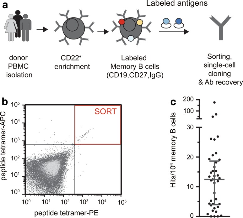
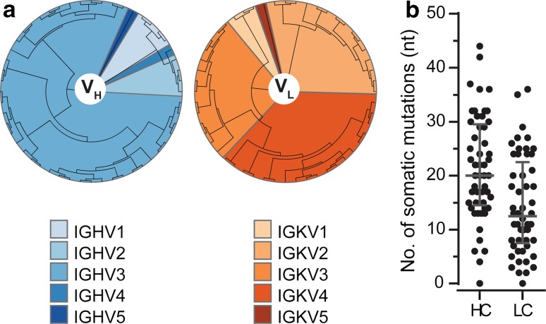
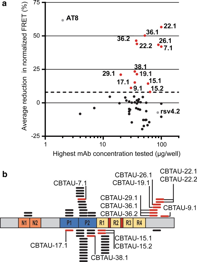
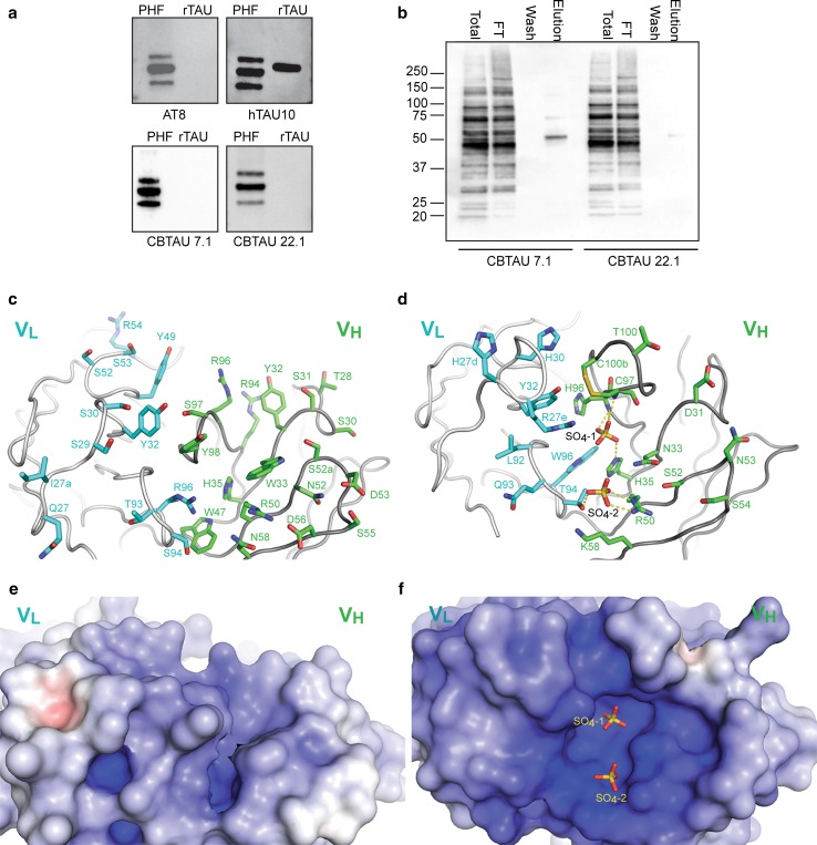
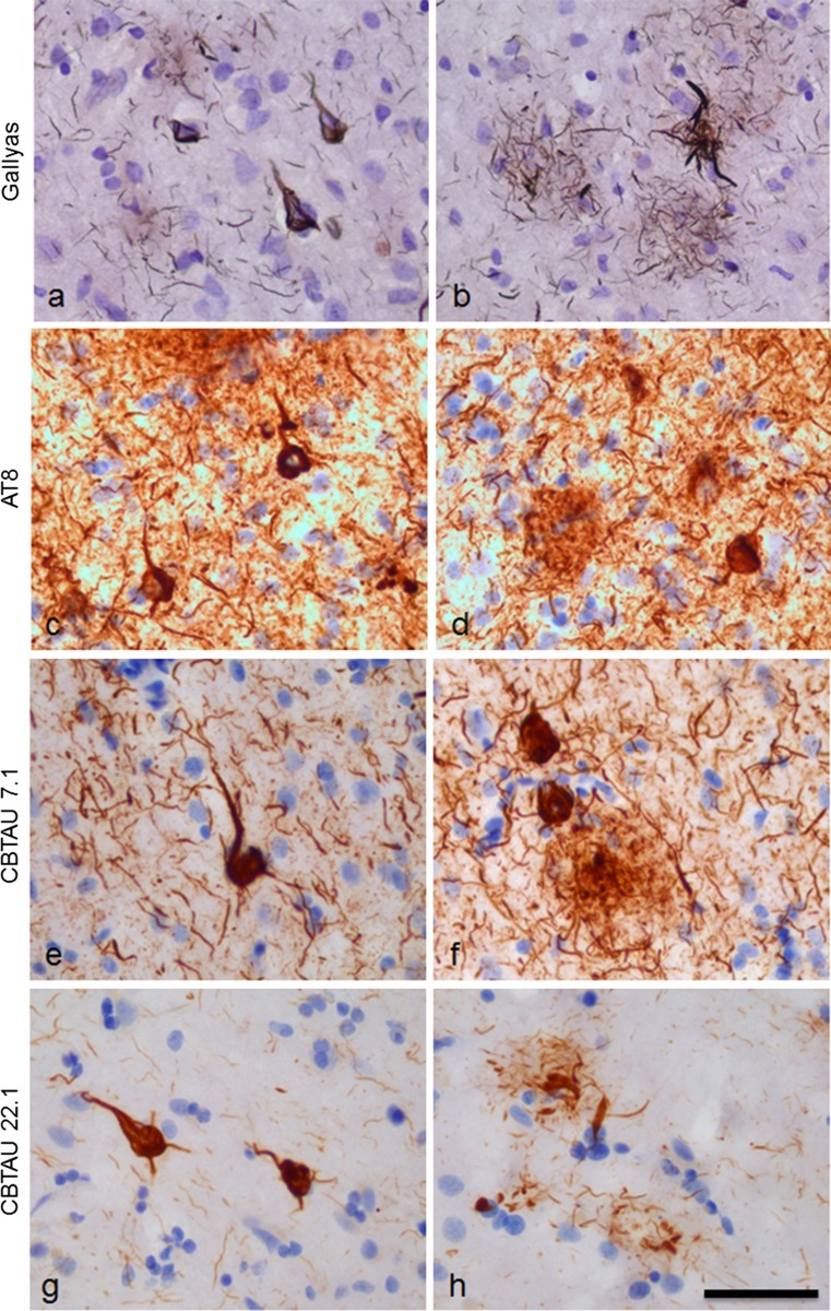
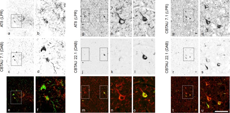
Several reports have described the presence of antibodies against Alzheimer's disease-associated hyperphosphorylated forms of tau in serum of healthy individuals. To characterize the specificities that can be found, we interrogated peripheral IgG+ memory B cells from asymptomatic blood donors for reactivity to a panel of phosphorylated tau peptides using a single-cell screening assay. Antibody sequences were recovered, cloned, and expressed as full-length IgGs. In total, 52 somatically mutated tau-binding antibodies were identified, corresponding to 35 unique clonal families. Forty-one of these antibodies recognize epitopes in the proline-rich and C-terminal domains, and binding of 26 of these antibodies is strictly phosphorylation dependent. Thirteen antibodies showed inhibitory activity in a P301S lysate seeded in vitro tau aggregation assay. Two such antibodies, CBTAU-7.1 and CBTAU-22.1, which bind to the proline-rich and C-terminal regions of tau, respectively, were characterized in more detail. CBTAU-7.1 recognizes an epitope that is similar to that of murine anti-PHF antibody AT8, but has different phospho requirements. Both CBTAU-7.1 and CBTAU-22.1 detect pathological tau deposits in post-mortem brain tissue. CBTAU-7.1 reveals a similar IHC distribution pattern as AT8, immunostaining (pre)tangles, threads, and neuritic plaques. CBTAU-22.1 shows selective detection of neurofibrillary changes by IHC. Taken together, these results suggest the presence of an ongoing antigen-driven immune response against tau in healthy individuals. The wide range of specificities to tau suggests that the human immune repertoire may contain antibodies that can serve as biomarkers or be exploited for therapy.

Keywords: Alzheimer’s disease; Memory B cell; Monoclonal antibody; Tau protein.


Conflict of interest statement

This work was fully funded by Janssen. A patent application relating to the CBTAU antibodies has been filed.

Figures

Fig. 1
Fig. 1
Recovery of naturally occurring monoclonal antibodies to pathogenic tau from asymptomatic individuals. a BSelex method used to recover tau-specific memory B cells. PBMCs were prepared from asymptomatic (non-AD) blood bank donors, and mature CD22+ B cells were positively selected with magnetic beads. Viable cells were stained with IgG-FITC, CD19-PerCPCy5.5, and CD27-PECy7, and with labeled peptide antigens and single-cell sorted on a Beckman Coulter MoFlo XDP. Antibody heavy and light variable chain sequences were recovered from single cells, cloned and expressed as full-length IgGs. b Polyvalent displays of tau phosphopeptides were labeled with APC or PE and gated on the double-positive population (PE+, APC+) to increase the signal-to-noise ratio. Memory B cells that showed reactivity to both APC- and PE-labeled peptides simultaneously were sorted as single cells into 96-well plates for further processing and recovery of IgH and IgL genes. Shown is a representative flow cytometric graph. c Estimated frequency of tau-specific B cells (Hits) per 106 memory B cells interrogated in the different sorts. Based on sorting losses (gated cells not being deposited), cloning efficiency (ability to recover both antibody heavy and light chains from single cells), and reconfirmation rate (whether the cloned antibody showed tau binding), we estimate that on average there were 12.5 potential hits per 1x10⁶ CD22+ CD19+CD27+IgG+ memory B cells examined across all sorts. The horizontal line indicates median, and error bars the inter-quartile range
Fig. 2
Fig. 2
Sequence analysis of recovered anti-tau monoclonal antibodies. a Phylogenetic analysis of recovered tau antibody heavy and light chain variable regions was performed using the neighbor-joining algorithm (Jukes Cantor model) and illustrated as a circular tree. b Number of somatic mutations in VH and VL genes in 52 antibodies analyzed from IgG+ memory B cells with reactivity to tau. Mutations and identification of the closest germline were determined using IgBlast and IMGT databases. The horizontal line indicates median and error bars the inter-quartile range
Fig. 3
Fig. 3
Antibody activity in an in vitro tau aggregation assay in relation to binding region and phospho-dependency. a Reduction in FRET signal of the highest tested concentration for each antibody. The names of antibodies that showed above-cutoff activity are indicated. Murine anti-tau antibody AT8 and anti-RSV-G antibody, RSV4.2, were included as positive and negative controls, respectively. Dotted line indicates a threshold (0% reduction + 2 × SD) below which measured activity is considered background. b Schematic showing the relative positions along tau isoform 2N4R of the tau peptides used to recover each of the 52 anti-tau antibodies. Highlighted (red) are the peptides recognized by those antibodies that show activity in the aggregation assay and the names of the respective antibodies are indicated. Cognate peptides of antibodies whose binding is dependent on phosphorylation are depicted above the representation of the tau isoform, while those of antibodies that bind to both phosphorylated and unphosphorylated peptides are depicted below the representation of the tau isoform. N1 and N2 indicate acidic inserts, P1 and P2 indicate proline-rich domains, and R1R4 indicate microtubule-binding repeat domains
Fig. 4
Fig. 4
Specificity and structural characterization of human anti-tau antibodies. a Reactivity of AT8 (phospho-dependent), htau10 (non-phospho–dependent), CBTAU-7.1 and CBTAU-22.1 to PHF and recombinant human tau-441 by Western blot. Triple bands characteristic of PHF-tau correspond to approximately 68, 64, and 60 kDa. b Western blot of flow-through (FT), wash (Wash) and eluate (Elution) fractions of immunoprecipitation (IP) of P301S transgenic mice brainstem homogenate with CBTAU-7.1 (left) and CBTAU-22.1 (right), as well as untreated P301S (Total) homogenate. The blot was stained with total-Tau antibody HT7. Total volumes of all samples were kept similar to enable comparison of concentrations. (c and d) Crystal structures of the combining sites of CBTAU-7.1 and CBTAU-22.1 Fab, respectively. Some solvent-exposed CDR residues in and around the antibody combining site are shown in cyan carbon atoms (light chain) and green carbon atoms (heavy chain). Two sulfates were modeled in the combining site of CBTAU-22.1 (d). (e and f) Electrostatic potential surfaces in and around the combining sites of CBTAU-7.1 Fab and CBTAU-22.1 Fab, respectively. Electrostatic surface potentials were calculated using the APBS program [5]. Negatively charged regions are red, positively charged regions are blue, and neutral regions are white (−15 to 15 K b T/e c potential range). Both CBTAU-7.1 and CBTAU-22.1 Fab exhibit highly positively charged surfaces in their combining sites
Fig. 5
Fig. 5
Overview of the pathological structures detected with Gallyas staining, AT8, CBTAU-7.1 and CBTAU-22.1. Staining was performed on 30 μm-thick, formalin-fixed, free-floating sections (temporal cortex, Alzheimer’s disease case, Braak VI). a, b Gallyas staining showing neurofibrillary changes. c, d AT8 immunostaining (0.25 μg/ml); e, f Immunostaining with CBTAU-7.1 (5 μg/ml). g, b Immunostaining with CBTAU-22.1 (5 μg/ml). Immunohistochemical detection with DAB (brown) and nuclei counterstained with haematoxylin (blue). Bar represents 50 μm
Fig. 6
Fig. 6
Double immunostaining with AT8, CBTAU-7.1 and CBTAU-22.1. Double immunostaining was performed on 5 μm-thick slide-mounted formalin-fixed, paraffin-embedded sections (temporal cortex, Alzheimer’s disease case, Braak VI). Double staining was analyzed with a spectral camera and digitally unmixed. Single staining patterns are presented separately (gray scale) and merged in artificial fluorescent colors. (af) Double immunostaining with AT8 (0.25 μg/ml, chromogen liquid permanent red, LPR) and CBTAU-7.1 (2.5 μg/ml, chromogen DAB). Areas indicated with dotted lines in a, c, and e are shown in higher magnification in b, d, and f respectively. (go) Double immunostaining with AT8 (0.25 μg/ml, LPR) and CBTAU-22.1 (10 μg/ml DAB). Areas indicated with dotted lines in g, j, and m are shown in higher magnification in h/i, k/l, and n/o respectively. (pu); Double immunostaining with CBTAU-7.1 (2.5 μg/ml, LPR) and CBTAU-22.1 (10 μg/ml, DAB). Areas indicated with dotted lines in p, r, and t are shown in higher magnification in q, s, and u respectively. Bar represents 100 μm in overviews and 50 μm in magnifications

References

    1. Afonine PV, Grosse-Kunstleve RW, Echols N, Headd JJ, Moriarty NW, Mustyakimov M, Terwilliger TC, Urzhumtsev A, Zwart PH, Adams PD. Towards automated crystallographic structure refinement with phenix.refine. Acta Crystallogr D Biol Crystallogr. 2012;68:352–367. doi: 10.1107/S0907444912001308. - DOI - PMC - PubMed
    1. Alafuzoff I, Pikkarainen M, Neumann M, Arzberger T, Al-Sarraj S, Bodi I, Bogdanovic N, Bugiani O, Ferrer I, Gelpi E, Gentleman S, Giaccone G, Graeber MB, Hortobagyi T, Ince PG, Ironside JW, Kavantzas N, King A, Korkolopoulou P, Kovacs GG, Meyronet D, Monoranu C, Nilsson T, Parchi P, Patsouris E, Revesz T, Roggendorf W, Rozemuller A, Seilhean D, Streichenberger N, Thal DR, Wharton SB, Kretzschmar H. Neuropathological assessments of the pathology in frontotemporal lobar degeneration with TDP43-positive inclusions: an inter-laboratory study by the BrainNet Europe consortium. J Neural Transm. 2015;122:957–972. doi: 10.1007/s00702-014-1304-1. - DOI - PubMed
    1. Allen B, Ingram E, Takao M, Smith MJ, Jakes R, Virdee K, Yoshida H, Holzer M, Craxton M, Emson PC, Atzori C, Migheli A, Crowther RA, Ghetti B, Spillantini MG, Goedert M. Abundant tau filaments and nonapoptotic neurodegeneration in transgenic mice expressing human P301S tau protein. J Neurosci. 2002;22:9340–9351. - PMC - PubMed
    1. Augustinack JC, Schneider A, Mandelkow EM, Hyman BT. Specific tau phosphorylation sites correlate with severity of neuronal cytopathology in Alzheimer’s disease. Acta Neuropathol. 2002;103:26–35. doi: 10.1007/s004010100423. - DOI - PubMed
    1. Baker NA, Sept D, Joseph S, Holst MJ, McCammon JA. Electrostatics of nanosystems: application to microtubules and the ribosome. Proc Natl Acad Sci USA. 2001;98:10037–10041. doi: 10.1073/pnas.181342398. - DOI - PMC - PubMed

Publication types