

Ritonavir and ixazomib kill bladder cancer cells by causing ubiquitinated protein accumulation
- PMID: 28342223
- PMCID: PMC5480085
- DOI: 10.1111/cas.13242
Ritonavir and ixazomib kill bladder cancer cells by causing ubiquitinated protein accumulation
Abstract
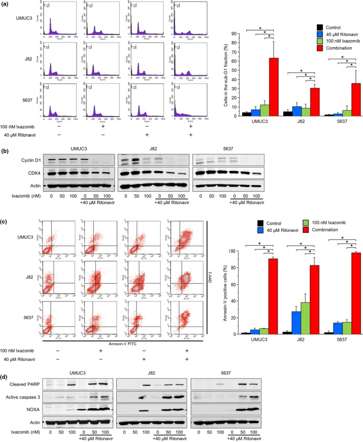
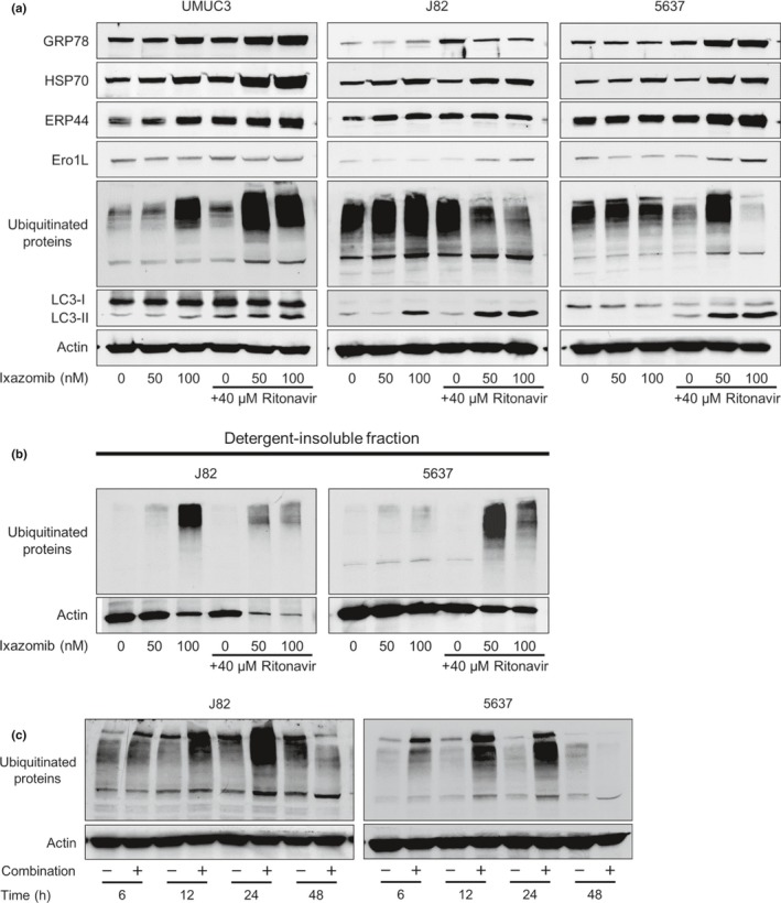
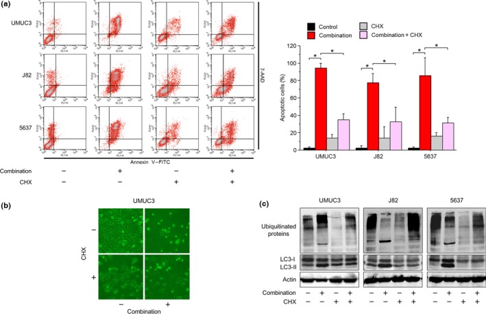
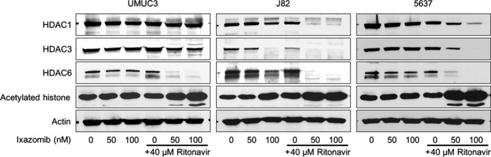
There is no curative treatment for advanced bladder cancer. Causing ubiquitinated protein accumulation and endoplasmic reticulum stress is a novel approach to cancer treatment. The HIV protease inhibitor ritonavir has been reported to suppress heat shock protein 90 and increase the amount of unfolded proteins in the cell. If the proteasome functions normally, however, they are rapidly degraded. We postulated that the novel proteasome inhibitor ixazomib combined with ritonavir would kill bladder cancer cells effectively by inhibiting degradation of these unfolded proteins and thereby causing ubiquitinated proteins to accumulate. The combination of ritonavir and ixazomib induced drastic apoptosis and inhibited the growth of bladder cancer cells synergistically. The combination decreased the expression of cyclin D1 and cyclin-dependent kinase 4, and increased the sub-G1 fraction significantly. Mechanistically, the combination caused ubiquitinated protein accumulation and endoplasmic reticulum stress. The combination-induced apoptosis was markedly attenuated by the protein synthesis inhibitor cycloheximide, suggesting that the accumulation of ubiquitinated proteins played an important role in the combination's antineoplastic activity. Furthermore, the combination induced histone acetylation cooperatively and the decreased expression of histone deacetylases was thought to be one mechanism of this histone acetylation. The present study provides a theoretical basis for future development of novel ubiquitinated-protein-accumulation-based therapies effective against bladder cancer.
Keywords: Bladder cancer; drug combinations; ixazomib; ritonavir; ubiquitinated proteins.
© 2017 The Authors. Cancer Science published by John Wiley & Sons Australia, Ltd on behalf of Japanese Cancer Association.
Figures

Similar articles
-
The Dual Histone Deacetylase-Proteasome Inhibitor RTS-V5 Acts Synergistically With Ritonavir to Induce Endoplasmic Reticulum Stress in Bladder Cancer Cells.Anticancer Res. 2021 Dec;41(12):5987-5996. doi: 10.21873/anticanres.15417. Anticancer Res. 2021. PMID: 34848452
-
Nelfinavir and Ritonavir Kill Bladder Cancer Cells Synergistically by Inducing Endoplasmic Reticulum Stress.Oncol Res. 2018 Mar 5;26(2):323-332. doi: 10.3727/096504017X14957929842972. Epub 2017 May 26. Oncol Res. 2018. PMID: 28560953 Free PMC article.
-
Ritonavir interacts with bortezomib to enhance protein ubiquitination and histone acetylation synergistically in renal cancer cells.Urology. 2012 Apr;79(4):966.e13-21. doi: 10.1016/j.urology.2011.11.033. Epub 2012 Jan 30. Urology. 2012. PMID: 22296623
-
[Pharmacological characteristics and clinical study results of the oral proteasome inhibitor ixazomib (NINLARO® capsules; 2.3 mg, 3 mg, and 4 mg)].Nihon Yakurigaku Zasshi. 2018;151(4):166-178. doi: 10.1254/fpj.151.166. Nihon Yakurigaku Zasshi. 2018. PMID: 29628465 Review. Japanese.
-
The human immunodeficiency virus protease inhibitor ritonavir is potentially active against urological malignancies.Onco Targets Ther. 2015 Apr 8;8:761-8. doi: 10.2147/OTT.S79776. eCollection 2015. Onco Targets Ther. 2015. PMID: 25914545 Free PMC article. Review.
Cited by
-
Fluvastatin potentiates anticancer activity of vorinostat in renal cancer cells.Cancer Sci. 2020 Jan;111(1):112-126. doi: 10.1111/cas.14225. Epub 2019 Nov 25. Cancer Sci. 2020. PMID: 31675763 Free PMC article.
-
Enhancing drug and cell line representations via contrastive learning for improved anti-cancer drug prioritization.NPJ Precis Oncol. 2024 May 18;8(1):106. doi: 10.1038/s41698-024-00589-8. NPJ Precis Oncol. 2024. PMID: 38762647 Free PMC article.
-
Repurposing old drugs as new inhibitors of the ubiquitin-proteasome pathway for cancer treatment.Semin Cancer Biol. 2021 Jan;68:105-122. doi: 10.1016/j.semcancer.2019.12.013. Epub 2019 Dec 26. Semin Cancer Biol. 2021. PMID: 31883910 Free PMC article. Review.
-
Ubiquitination-Related Molecular Subtypes and a Novel Prognostic Index for Bladder Cancer Patients.Pathol Oncol Res. 2021 Oct 29;27:1609941. doi: 10.3389/pore.2021.1609941. eCollection 2021. Pathol Oncol Res. 2021. PMID: 34776794 Free PMC article.
-
Ritonavir's Evolving Role: A Journey from Antiretroviral Therapy to Broader Medical Applications.Curr Oncol. 2024 Oct 8;31(10):6032-6049. doi: 10.3390/curroncol31100450. Curr Oncol. 2024. PMID: 39451754 Free PMC article. Review.
References
-
- von der Maase H, Sengelov L, Roberts JT et al Long‐term survival results of a randomized trial comparing gemcitabine plus cisplatin, with methotrexate, vinblastine, doxorubicin, plus cisplatin in patients with bladder cancer. J Clin Oncol 2005; 23: 4602–8. - PubMed
-
- Naujokat C, Hoffmann S. Role and function of the 26S proteasome in proliferation and apoptosis. Lab Invest 2002; 82: 965–80. - PubMed
-
- Mimnaugh EG, Xu W, Vos M et al Simultaneous inhibition of hsp 90 and the proteasome promotes protein ubiquitination, causes endoplasmic reticulum‐derived cytosolic vacuolization, and enhances antitumor activity. Mol Cancer Ther 2004; 3: 551–66. - PubMed
MeSH terms
Substances
LinkOut - more resources
Full Text Sources
Other Literature Sources
Medical
Molecular Biology Databases
Research Materials
