

Contributions of intestinal epithelial barriers to health and disease
- PMID: 28342899
- PMCID: PMC5958612
- DOI: 10.1016/j.yexcr.2017.03.036
Contributions of intestinal epithelial barriers to health and disease
Abstract
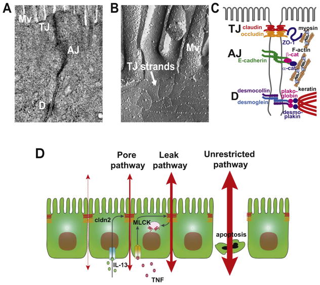
A core function of epithelia is to form barriers that separate tissue compartments within complex organisms. These barriers also separate the internal milieu from the external environment and are, therefore, an essential component of host defense. However, in many cases, a perfect barrier would be improbable with life itself. Examples include the air spaces within the lungs, the renal tubules, and the intestines. Here, we focus on the mechanisms by which barriers are assembled, maintained, and regulated in the context of health and disease. Because of its unique challenges and extensive study, we focus on the gastrointestinal tract as an organ-specific example of the essential contributions of the paracellular barrier to life.
Copyright © 2017. Published by Elsevier Inc.
Figures
References
-
- Marchiando AM, Graham WV, Turner JR. Epithelial barriers in homeostasis and disease. Annu Rev Pathol. 2010;5:119–144. - PubMed
-
- Meddings JB, Westergaard H. Intestinal glucose transport using perfused rat jejunum in vivo: model analysis and derivation of corrected kinetic constants. Clin Sci. 1989;76:403–413. - PubMed
-
- Turner JR, Rill BK, Carlson SL, Carnes D, Kerner R, Mrsny RJ, Madara JL. Physiological regulation of epithelial tight junctions is associated with myosin light-chain phosphorylation. Am J Physiol. 1997;273:C1378–C1385. - PubMed
-
- Pappenheimer JR, Reiss KZ. Contribution of solvent drag through intercellular junctions to absorption of nutrients by the small intestine of the rat. J Membr Biol. 1987;100:123–136. - PubMed
Publication types
MeSH terms
Grants and funding
LinkOut - more resources
Full Text Sources
Other Literature Sources
