. 2017 Jun;46(6):785-793.
doi: 10.1007/s00256-017-2616-7. Epub 2017 Mar 25.

Skeletal development of the hand and wrist: digital bone age companion-a suitable alternative to the Greulich and Pyle atlas for bone age assessment?

Affiliations

Skeletal development of the hand and wrist: digital bone age companion-a suitable alternative to the Greulich and Pyle atlas for bone age assessment?

Paul M Bunch et al. Skeletal Radiol. 2017 Jun.

Abstract

Purpose: To assess reader performance and subjective workflow experience when reporting bone age studies with a digital bone age reference as compared to the Greulich and Pyle atlas (G&P). We hypothesized that pediatric radiologists would achieve equivalent results with each method while digital workflow would improve speed, experience, and reporting quality.

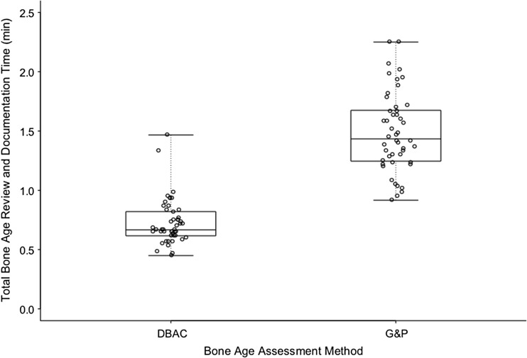
Materials and methods: IRB approval was obtained for this HIPAA-compliant study. Two pediatric radiologists performed research interpretations of bone age studies randomized to either the digital (Digital Bone Age Companion, Oxford University Press) or G&P method, generating reports to mimic clinical workflow. Bone age standard selection, interpretation-reporting time, and user preferences were recorded. Reports were reviewed for typographical or speech recognition errors. Comparisons of agreement were conducted by way of Fisher's exact tests. Interpretation-reporting times were analyzed on the natural logarithmic scale via a linear mixed model and transformed to the geometric mean. Subjective workflow experience was compared with an exact binomial test. Report errors were compared via a paired random permutation test.

Results: There was no difference in bone age determination between atlases (p = 0.495). The interpretation-reporting time (p < 0.001) was significantly faster with the digital method. The faculty indicated preference for the digital atlas (p < 0.001). Signed reports had fewer errors with the digital atlas (p < 0.001).

Conclusions: Bone age study interpretations performed with the digital method were similar to those performed with the Greulich and Pyle atlas. The digital atlas saved time, improved workflow experience, and reduced reporting errors relative to the Greulich and Pyle atlas when integrated into electronic workflow.

Keywords: Bone age; Children; Development; Radiography; Skeletal age; Skeletal maturity.


Conflict of interest statement

Disclosures

Cree Gaskin receives author royalties from Thieme Medical Publishers and Oxford University Press, as well as research grant funding from Carestream Health. This author did not participate in bone age study interpretation or in the statistical analysis.

Drs. Bunch, Altes, and McIlhenny and Mr. Patrie have no financial disclosures.

Ethics approval

All procedures performed in studies involving human participants were in accordance with the ethical standards of the institutional research committee and with the 1964 Helsinki Declaration and its later amendments. For this type of study formal consent is not required.

Conflict of interest/disclosure statement

The corresponding author receives author royalties from Oxford University Press and Thieme Medical Publishers. Additionally, the corresponding author has a research grant from Carestream Health. This author did not participate in bone age study interpretation or in the statistical analysis. The other authors have no financial disclosures.

Figures

Fig. 1
Fig. 1
Demonstration of bone age study interpretation with the digital method integrated into clinical workflow. (The display of patient information in the software is from a hypothetical patient for illustrative purposes. Any resemblance to that of an actual patient is coincident.) Figure 1b–e from Skeletal Development of the Hand and Wrist: Digital Bone Age Companion by Gaskin et al, 2011. By permission of Oxford University Press. a Screenshot from RIS-EHR demonstrates anonymized bone age study requisition and “BoneAge” button (orange arrow) in the graphical user interface. Clicking the button launches bone age software and initiates XML file drop containing the patient’s date of birth, date of study, and gender. b Screenshot of bone age software. The software references the XML file and then displays the bone age standard closest to the patient’s age and matching the patient’s gender for the most likely match in the typical patient. Users may zoom in on skeletal features and adjust the window level and width to their preference. c Screenshot of bone age software demonstrates optional annotated standards to aid the interpreter in choosing the best match. Up to three standards (only two shown) may be reviewed side-by-side to further aid decision making. Clicking the calculator button (calculator icon at bottom left) sends the patient’s date of birth, date of study, gender, and chosen standard to the calculator. d Screenshot of bone age calculator. The calculator uses the correct standard deviation value to perform the skeletal maturity calculation. The software includes settings that allow for adjustments in how the calculation is performed to accommodate regional practice differences. The user can manually edit the estimated skeletal age if it falls between two standards. Clicking the “Generate Report” button (bottom right) creates a structured report that can be copy/pasted to the reporting system for final review and signature. e Example of structured report generated by bone age software based upon user-chosen bone age standard and automatically imported patient demographics from the RIS-EHR or PACS
Fig. 2
Fig. 2
Box and whisker plot demonstrating the distribution of the bone age interpretation-report cycle times in minutes between the integrated electronic method of the Digital Bone Age Companion (DBAC) and the method of Greulich and Pyle (G&P) for the two faculty pediatric radiologists combined. Interpretation-report cycle time was defined as the time interval beginning with the study loading on PACS and ending with signing of the corrected report. Each circle represents an interpretation-report cycle time; the boxes represent the middle 50%; the lines within the boxes represent the mean; the superior- and inferior-most lines represent the maximum and minimum times, respectively

References

    1. Martin DD, Wit JM, Z ’ev H. The use of bone age in clinical practice—part 1. Horm Res Pædiatr. 2011;76:1–9. doi: 10.1159/000329372. - DOI - PubMed
    1. Lynnerup N, Belard E, Buch-Olsen K. Intra- and interobserver error of the Greulich-Pyle method as used on a Danish forensic sample. Forensic Sci Int. 2008;179:242.e1–6. doi: 10.1016/j.forsciint.2008.05.005. - DOI - PubMed
    1. Mentzel H-J, Vilser C, Eulenstein M, et al. Assessment of skeletal age at the wrist in children with a new ultrasound device. Pediatr Radiol. 2005;35:429–33. doi: 10.1007/s00247-004-1385-3. - DOI - PubMed
    1. Tanner JM, Healy M, Goldstein H, Cameron N. Assessment of skeletal maturity and prediction of adult height (TW3 method) London; New York: W.B. Saunders; 2001.
    1. Thodberg HH, Kreiborg S, Juul A, Pedersen KD. The BoneXpert method for automated determination of skeletal maturity. IEEE Trans Med Imaging. 2009;28:52–66. doi: 10.1109/TMI.2008.926067. - DOI - PubMed

LinkOut - more resources