. 2017 Nov;242(17):1643-1656.
doi: 10.1177/1535370217701006. Epub 2017 Mar 26.

Toward improved myocardial maturity in an organ-on-chip platform with immature cardiac myocytes

Affiliations

Toward improved myocardial maturity in an organ-on-chip platform with immature cardiac myocytes

Sean P Sheehy et al. Exp Biol Med (Maywood). 2017 Nov.

Abstract

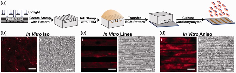
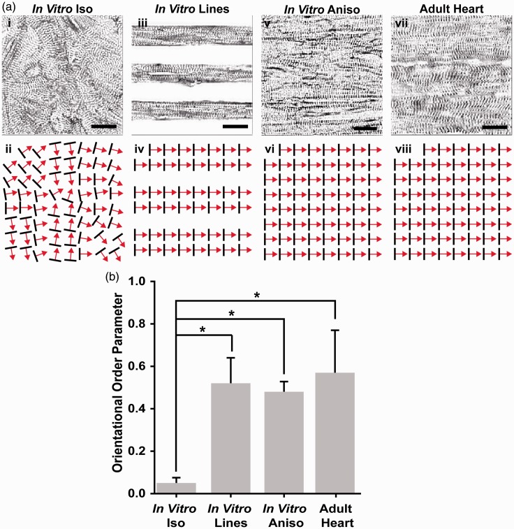
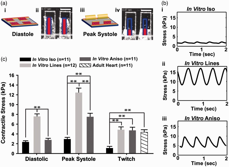
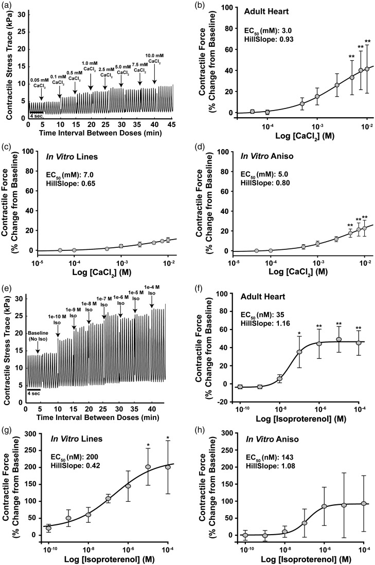
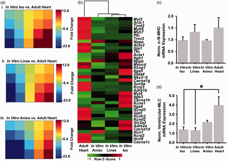
In vitro studies of cardiac physiology and drug response have traditionally been performed on individual isolated cardiomyocytes or isotropic monolayers of cells that may not mimic desired physiological traits of the laminar adult myocardium. Recent studies have reported a number of advances to Heart-on-a-Chip platforms for the fabrication of more sophisticated engineered myocardium, but cardiomyocyte immaturity remains a challenge. In the anisotropic musculature of the heart, interactions between cardiac myocytes, the extracellular matrix (ECM), and neighboring cells give rise to changes in cell shape and tissue architecture that have been implicated in both development and disease. We hypothesized that engineered myocardium fabricated from cardiac myocytes cultured in vitro could mimic the physiological characteristics and gene expression profile of adult heart muscle. To test this hypothesis, we fabricated engineered myocardium comprised of neonatal rat ventricular myocytes with laminar architectures reminiscent of that observed in the mature heart and compared their sarcomere organization, contractile performance characteristics, and cardiac gene expression profile to that of isolated adult rat ventricular muscle strips. We found that anisotropic engineered myocardium demonstrated a similar degree of global sarcomere alignment, contractile stress output, and inotropic concentration-response to the β-adrenergic agonist isoproterenol. Moreover, the anisotropic engineered myocardium exhibited comparable myofibril related gene expression to muscle strips isolated from adult rat ventricular tissue. These results suggest that tissue architecture serves an important developmental cue for building in vitro model systems of the myocardium that could potentially recapitulate the physiological characteristics of the adult heart. Impact statement With the recent focus on developing in vitro Organ-on-Chip platforms that recapitulate tissue and organ-level physiology using immature cells derived from stem cell sources, there is a strong need to assess the ability of these engineered tissues to adopt a mature phenotype. In the present study, we compared and contrasted engineered tissues fabricated from neonatal rat ventricular myocytes in a Heart-on-a-Chip platform to ventricular muscle strips isolated from adult rats. The results of this study support the notion that engineered tissues fabricated from immature cells have the potential to mimic mature tissues in an Organ-on-Chip platform.

Keywords: Heart-on-a-Chip; Muscular thin films; cardiac contractility; cardiac tissue engineering.


Figures

Figure 1
Figure 1
Controlling the tissue architecture of engineered myocardium using ECM guidance cues. (a) The architecture of engineered cardiac tissues was controlled by micro-contact printing the ECM protein fibronectin (FN) into the desired pattern. Three distinct ECM patterns were chosen for this study to assess the contribution of tissue architecture to the maturation of engineered myocardium in vitro. (b) In Vitro Iso: coverslips coated uniformly with FN (i), gave rise to monolayers of randomly oriented cardiomyocytes (ii). (c) In Vitro Lines: micro-contact printed 15 µm wide FN lines spaced 15 µm apart (i) produced linear arrays of highly aligned cardiac myocytes (ii). (d) In Vitro Aniso: micro-contact printed 15 µm wide FN lines spaced 2 µm apart (i) produced confluent anisotropic sheets of cardiac myocytes (ii). For panels bi, ci, di, scale bars = 10 µm. For panels bii, cii, dii, scale bars = 100 µm
Figure 2
Figure 2
Comparison of global sarcomere alignment in engineered and adult myocardium. (a) Assessment of sarcomeric α-actinin fluorescence micrographs of isotropic samples (i) revealed random z-line organization as illustrated in the schematic below (ii). In contrast, α-actinin micrographs of cardiac myocytes cultured on 15 µm wide FN lines spaced 15 µm apart (iii), displayed the high degree of parallel alignment expected from the ECM patterning, as illustrated in this schematic (iv). The z-lines of cardiomyocytes cultured on 15 µm wide FN lines spaced 2 µm apart (v) also displayed the degree of parallel z-line alignment expected from this ECM pattern, as illustrated in this schematic (vi). Comparison of micro-contact printed engineered myocardium to the z-line organization observed in histological sections of the adult rat ventricular myocardium (vii) reveals a similar level of global sarcomere alignment as illustrated in this schematic (viii). (b) Statistical comparison of global sarcomere alignment quantified using the Orientational Order Parameter revealed that the anisotropic engineered myocardium showed similar levels of alignment to the adult rat ventricular myocardium. n = 3 tissues for In Vitro Iso, In Vitro Lines, and In Vitro Aniso; n = 4 ventricles for Adult Heart. Scale bars = 10 µm. *= P < 0.05 versus In vitro Iso
Figure 3
Figure 3
Measurement and comparison of contractile performance in engineered rat myocardium. (a) Engineered myocardium cultured on MTF cantilevers lay almost flat against the substrate during diastole (i, ii) and curled up out of the plane of the substrate during systolic contraction (iii, iv), scale bars = 1 mm. (b) High speed video recording allows calculation of stress traces during contraction cycles. Representative stress traces for engineered tissues In Vitro Iso (i), In Vitro Lines (ii), In Vitro Aniso (iii). (c) Statistical comparison of diastolic (rest), peak systolic (maximum contraction), and twitch (difference between diastolic and peak systolic) stresses generated by engineered myocardium with isotropic (In Vitro Iso) and anisotropic (In Vitro Lines, In Vitro Aniso) sarcomere organization. Twitch stress from baseline measurements of adult rat muscle strip contractility (Adult Heart) were calculated and compared to values calculated for engineered myocardium. In Vitro Lines, and In Vitro Aniso engineered myocardium demonstrated twitch stress values within the range reported for adult myocardium,, (In vitro Iso: n = 11 films, #chips = 3; In Vitro Lines: n = 12 films, #chips = 3; In Vitro Aniso: #chips = 3; n = 11 films, Adult Heart: n = 11 muscle strips), **=P<0.001 versus In vitro Iso
Figure 4
Figure 4
Measurement and comparison of inotropic response to CaCl2 and isoproterenol in engineered and adult rat myocardium. (a) Example stress trace from MTF contractility measurements illustrating the Calcium chloride (CaCl2) concentration response protocol used to compare the inotropic response profiles of engineered and adult rat myocardium. CaCl2 was administered cumulatively over 5 min intervals to achieve bath concentrations ranging from 0.05 to 10 mM, and contractile stress measurements were recorded at each concentration. (b) Percent change from baseline contractile response profile of ventricular muscle strips isolated from adult rats (Adult Heart) to increasing CaCl2 concentration, EC50 = 3 mM, n = 6 muscle strips. (c) Percent change from baseline contractile response profile of engineered myocardium comprised of neonate rat cardiac myocytes cultured on 15 µm wide FN lines spaced 15 µm apart (In Vitro Lines) to increasing CaCl2 concentration, EC50 = 7 mM, n = 9 MTFs, #chips = 2. (d) Percent change from baseline contractile response profile of engineered myocardium comprised of neonate rat cardiac myocytes cultured on 15 µm wide FN lines spaced 2 µm apart (In Vitro Aniso) to increasing CaCl2 concentration, EC50 = 5 nM, n = 15 MTFs, #chips = 3. (e) Example stress trace from MTF contractility measurements illustrating the Isoproterenol (Iso) concentration–response protocol used to compare the inotropic response profiles of engineered and adult rat myocardium. Baseline contractile stress measurements were recorded for 10 min, then Iso concentrations ranging from 1010 to 104 M were administered in 5 min intervals and recordings of contractile force were taken for each muscle construct. (f) Percent change from baseline contractile response profile of ventricular muscle strips isolated from adult rats (Adult Heart) to isoproterenol exposure, EC50 = 35 nM, n = 4 muscle strips. (g) Percent change from baseline contractile response profile of engineered myocardium comprised of neonate rat cardiac myocytes cultured on 15 µm wide FN lines spaced 15 µm apart (In Vitro Lines) to isoproterenol exposure, EC50 = 200 nM, n = 13 MTFs, #chips = 4. (h) Percent change from baseline contractile response profile of engineered myocardium comprised of neonate rat cardiac myocytes cultured on 15 µm wide FN lines spaced 2 µm apart (In Vitro Aniso) to Iso exposure, EC50 = 143 nM, n = 15 MTFs, #chips = 4. * = P < 0.05 versus baseline, ** = P < 0.001 versus baseline. Data presented as mean ± SEM
Figure 5
Figure 5
Comparison of gene expression profiles in engineered and adult rat myocardium. RT-qPCR measurements were made for a panel of genes associated with myocardial development (Supp. Table S1) on In Vitro Iso, In Vitro Lines, and In Vitro Aniso engineered myocardium, as well as explants from adult rat ventricular tissue. (a) Fold change values were calculated against the Adult Heart tissue for the In Vitro Iso (i), In Vitro Lines (ii), In Vitro Aniso (iii) tissues and analyzed with Gene Expression Dynamics Inspector (GEDI) to visualize global differences in the expression profiles between the engineered myocardium versus in vivo myocardium. (b) Heat map illustrating hierarchical clustering of the engineered and in vivo myocardial tissue based on mean 2−ΔCt expression values. (c) α-/β-myosin heavy chain ratio was not significantly different between engineered myocardium and adult myocardium, (d) In Vitro Iso and In Vitro Lines engineered myocardium samples both exhibited significant differences in ventricular myosin light chain (MYL2) expression from Adult Heart tissue explants, whereas the In Vitro Aniso samples did not. n = 3 tissues for all samples, * = P < 0.05 versus Adult Heart. Data presented as mean ± SEM

References

    1. Burrows MT. Rhythmical activity of isolated heart muscle cells in vitro. Science 1912; 36: 90–2. - PubMed
    1. Greek R, Menache A. Systematic reviews of animal models: methodology versus epistemology. Int J Med Sci 2013; 10: 206–21. - PMC - PubMed
    1. Eschenhagen T, Eder A, Vollert I, Hansen A. Physiological aspects of cardiac tissue engineering. Am J Physiol Heart Circ Physiol 2012; 303: H133–43. - PubMed
    1. Zimmermann WH, Schneiderbanger K, Schubert P, Didie M, Munzel F, Heubach JF, Kostin S, Neuhuber WL, Eschenhagen T. Tissue engineering of a differentiated cardiac muscle construct. Circ Res 2002; 90: 223–30. - PubMed
    1. Nawroth JC, Parker KK. Design standards for engineered tissues. Biotechnol Adv 2013; 31: 632–7. - PMC - PubMed

Publication types