. 2017 Apr 11;8(4):803-812.
doi: 10.1016/j.stemcr.2017.02.016. Epub 2017 Mar 23.

Inducible and Deterministic Forward Programming of Human Pluripotent Stem Cells into Neurons, Skeletal Myocytes, and Oligodendrocytes

Affiliations

Inducible and Deterministic Forward Programming of Human Pluripotent Stem Cells into Neurons, Skeletal Myocytes, and Oligodendrocytes

Matthias Pawlowski et al. Stem Cell Reports. .

Abstract

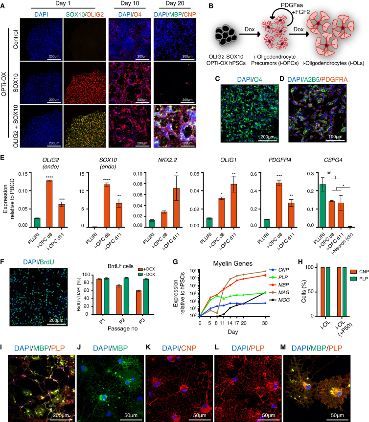
The isolation or in vitro derivation of many human cell types remains challenging and inefficient. Direct conversion of human pluripotent stem cells (hPSCs) by forced expression of transcription factors provides a potential alternative. However, deficient inducible gene expression in hPSCs has compromised efficiencies of forward programming approaches. We have systematically optimized inducible gene expression in hPSCs using a dual genomic safe harbor gene-targeting strategy. This approach provides a powerful platform for the generation of human cell types by forward programming. We report robust and deterministic reprogramming of hPSCs into neurons and functional skeletal myocytes. Finally, we present a forward programming strategy for rapid and highly efficient generation of human oligodendrocytes.

Keywords: human pluripotent stem cells; neurons; oligodendrocyte progenitor cells; reprogramming; skeletal myocytes.


Figures

None
Graphical abstract
Figure 1
Figure 1
Development of an Optimized Inducible Gene Overexpression System (A) Workflow for targeting the hROSA26 and AAVS1 loci with the Tet-ON system in hPSCs for inducible EGFP expression (i-EGFP). Cas9n, D10A nickase mutant Cas9 endonuclease; ZFN, zinc-finger nucleases; rtTA, reverse tetracycline transactivator; TRE, Tet-responsive element. (B) Schematic of the four outcomes following generation of dual GSH-targeted inducible EGFP hESCs: clonal lines were categorized based on the number of successfully targeted alleles of the hROSA26 and AAVS1 loci. (C) Detection of the rtTA protein by western blot in heterozygous (HET) and homozygous (HOM) hROSA26-CAG-rtTA hESCs. Homozygous targeting results in increased rtTA protein expression. hESCs with random integration of a second-generation rtTA (M2-rtTA) and wild-type hESCs are shown as positive and negative reference. α-Tubulin, loading control. (D) Median fluorescent intensity (MFI) of EGFP expression in the various dual GSH-targeted i-EGFP hESCs described in (B). Cells were analyzed by flow cytometry in non-induced conditions (CTR) or following 5 days of dox. AAVS1-CAG-EGFP and wild-type (WT) hESCs were included for comparison. Statistical analysis of dox-treated groups demonstrated that EGFP levels were highest in double-homozygous clones (each data point, n = 1–5, represents a clonal line; mean ± SEM; one-way ANOVA with post hoc Dunnett's test; ∗∗p < 0.01, ∗∗∗∗p < 0.0001). (E) Flow cytometry of EGFP OPTi-OX hESCs after 5 days of dox treatment. Non-induced cells were included as negative control. (F and G) EGFP induction and rescue kinetics (F) and dox dose-response (G) in EGFP OPTi-OX hESCs detected by flow cytometry (n = 2 biological replicates; mean ± SEM; all values normalized to the maximum fluorescence intensity after 5 days of dox). (H–K) Immunocytochemistry (ICC) for lineage-specific markers in undifferentiated EGFP OPTi-OX hESCs and following differentiation into the germ layers.
Figure 2
Figure 2
Forward Programming of hPSCs into Neurons (A) Experimental approach for conversion of NGN2 OPTi-OX hPSCs into i-Neurons. (B) Time course of i-Neuron generation from hESCs by qPCR demonstrating the expression pattern of pluripotency factors (OCT4 and NANOG), pan-neuronal (MAP2 and SYP), forebrain (BRN2, FOXG1), and glutamatergic neuronal marker genes (VGlut2, GRIA4) (n = 3 biological replicates; mean ± SEM; relative to PBGD and normalized to pluripotency). (C) Phase contrast images illustrating the morphological changes during i-Neuron generation (a corresponding time-lapse is shown in Movie S1). (D) ICC for the pan-neuronal marker proteins βIII-tubulin (TUBB3) and microtubule-associated protein 2 (MAP2) during the generation of i-Neurons. (E) Quantification of βIII-tubulin-positive neuronal cells by ICC after 1 week of induction. Undifferentiated cells were used as negative control, and numbers are reported for i-Neuron generation in newly isolated NGN2 OPTi-OX hESCs and after 25 passages. (F and G) ICC for neuronal markers in i-Neurons 14 days after induction.
Figure 3
Figure 3
Forward Programming of hPSCs into Skeletal Myocytes (A) Experimental approach for rapid single-step conversion of MYOD1 OPTi-OX hPSCs into skeletal myocytes (i-Myocytes) following treatment with dox and RA. (B) Representative ICC for MYOD1 before (CTR) and after induction with dox. This demonstrates homogeneous induction of transgene expression, paralleled by downregulation of the pluripotency factors NANOG and OCT4. (C) Effect of RA on myocyte forward programming compared with otherwise identical control (CTR) induction conditions (see Figure S2B for the entire signaling molecule screen). (D) qPCR of the temporal expression pattern of pluripotency factors (top panel) and myocyte marker genes during i-Myocyte generation (n = 3 biological replicates, mean ± SEM; relative to PBGD and normalized to pluripotency). (E and F) ICC for skeletal myocyte markers in i-Myocytes. (G and H) Quantification of MHC-positive cells by flow cytometry 10 days after induction. Undifferentiated cells were used as negative control, and figures are reported for i-Myocyte generation in newly isolated MYOD1 OPTi-OX hESCs, or in the same cells following 50 passages (+P50) (n = 3 biological replicates; mean ± SEM). (I) qPCR for total MYOD1, endogenous MYOD1, and MYOG 2 days post induction with different dox concentrations (n = 3 biological replicates; mean ± SEM). (J) ICC for myogenin and myosin heavy chain following 5 days of induction with different dox concentrations. Non-converted, proliferative cell clusters appeared when the dox concentration was lowered to 0.125 μg/mL. Further reduction of dox resulted in an increase in non-myocyte cell populations.
Figure 4
Figure 4
Forward Programming of hPSCs into Oligodendrocytes (A) ICC for inducible transgenes after 1 day of induction (left column), the OPC marker O4 after 10 days (middle), and the oligodendrocyte markers CNP and MBP after 20 days (right). (B) Experimental approach for rapid conversion of OLIG2-SOX10 OPTi-OX hPSCs into the oligodendrocyte lineage cells (i-OPCs and i-OLs). (C and D) Characterization of i-OPCs by ICC for OPC surface markers (A2B5, O4, PDGFRA). (E) Characterization of i-OPCs by qPCR compared with hPSCs (PLURI). As transcription of CSPG4 (NG2) was also detected in hPSCs, we included i-Neurons as negative control (n = 3 biological replicates; mean ± SEM; all values relative to PBGD; one-way ANOVA with post hoc Dunnett's test; p < 0.05; ∗∗p < 0.01, ∗∗∗p < 0.001, ∗∗∗∗p < 0.0001; ns, p > 0.05). (F) Immunostaining for BrdU (left panel) and quantification of BrdU-positive cells following three serial passages of i-OPCs every 4 days and concomitant BrdU-pulses (n = 3 biological replicates; mean ± SEM; P, passage). (G) qPCR of the temporal expression pattern of genes encoding for the myelin-associated proteins (CNP, MAG, MBP, MOG, and PLP) during i-OL generation. OLIG2-SOX10 OPTi-OX hPSCs were induced in oligodendrocyte medium supplemented with PDGFaa and FGF2. After 1 week of induction, mitogens were withdrawn to enable terminal differentiation (n = 3 biological replicates; mean ± SEM; all values relative to PBGD and normalized to pluripotency). (H) Quantification of CNP and PLP expressing i-OLs derived from OLIG2-SOX10 OPTi-OX hPSCs after 20 days of induction by ICC. Undifferentiated cells were used as negative control, and figures are reported for newly isolated OLIG2-SOX10 OPTi-OX hPSCs and after 50 passages (+P50). (I–M) ICC providing an overview (I) and high-magnifications (J–M) of mature pre-myelinating oligodendrocytes.

References

    1. Abujarour R., Bennett M., Valamehr B., Lee T.T., Robinson M., Robbins D., Le T., Lai K., Flynn P. Myogenic differentiation of muscular dystrophy-specific induced pluripotent stem cells for use in drug discovery. Stem Cells Transl. Med. 2014;3:149–160. - PMC - PubMed
    1. Albini S., Coutinho P., Malecova B., Giordani L., Savchenko A., Forcales S.V., Puri P.L. Epigenetic reprogramming of human embryonic stem cells into skeletal muscle cells and generation of contractile myospheres. Cell Rep. 2013;3:661–670. - PMC - PubMed
    1. Bar-Nur O., Brumbaugh J., Verheul C., Apostolou E., Pruteanu-Malinici I., Walsh R.M., Ramaswamy S., Hochedlinger K. Small molecules facilitate rapid and synchronous iPSC generation. Nat. Methods. 2014;11:1170–1176. - PMC - PubMed
    1. Baron U., Bujard H. Tet repressor-based system for regulated gene expression in eukaryotic cells: principles and advances. Methods Enzymol. 2000;327:401–421. - PubMed
    1. Bertero A., Pawlowski M., Ortmann D., Snijders K., Yiangou L., Cardoso de Brito M., Brown S., Bernard W.G., Cooper J.D., Giacomelli E. Optimized inducible shRNA and CRISPR/Cas9 platforms for in vitro studies of human development using hPSCs. Development. 2016;143:4405–4418. - PMC - PubMed

Publication types