. 2017 Apr 6;66(1):22-37.e9.
doi: 10.1016/j.molcel.2017.02.017. Epub 2017 Mar 23.

Circ-ZNF609 Is a Circular RNA that Can Be Translated and Functions in Myogenesis

Affiliations

Circ-ZNF609 Is a Circular RNA that Can Be Translated and Functions in Myogenesis

Ivano Legnini et al. Mol Cell. .

Abstract

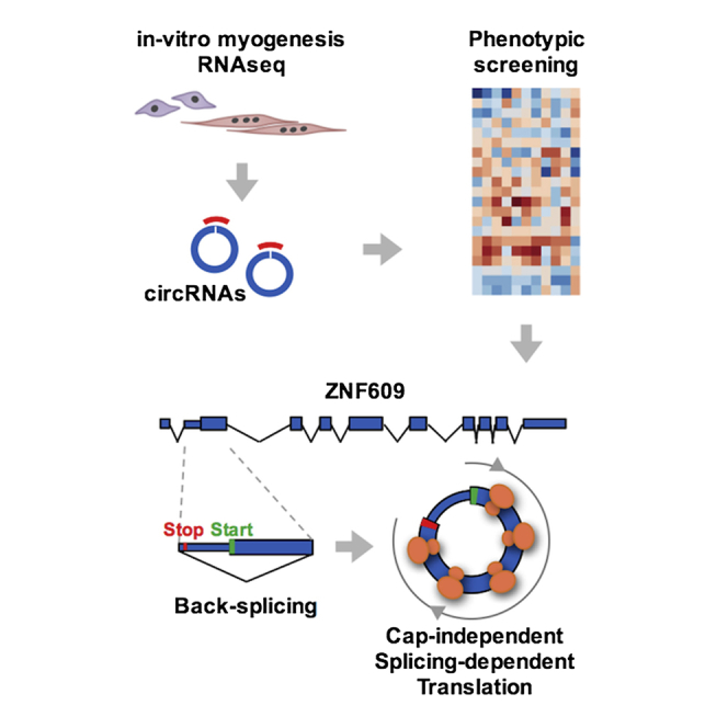
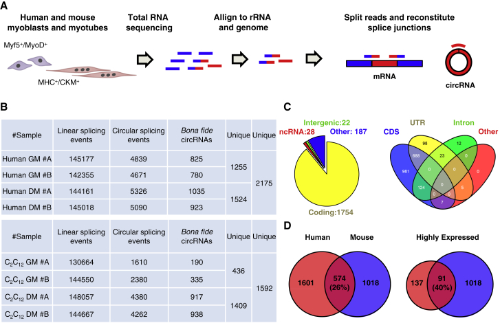
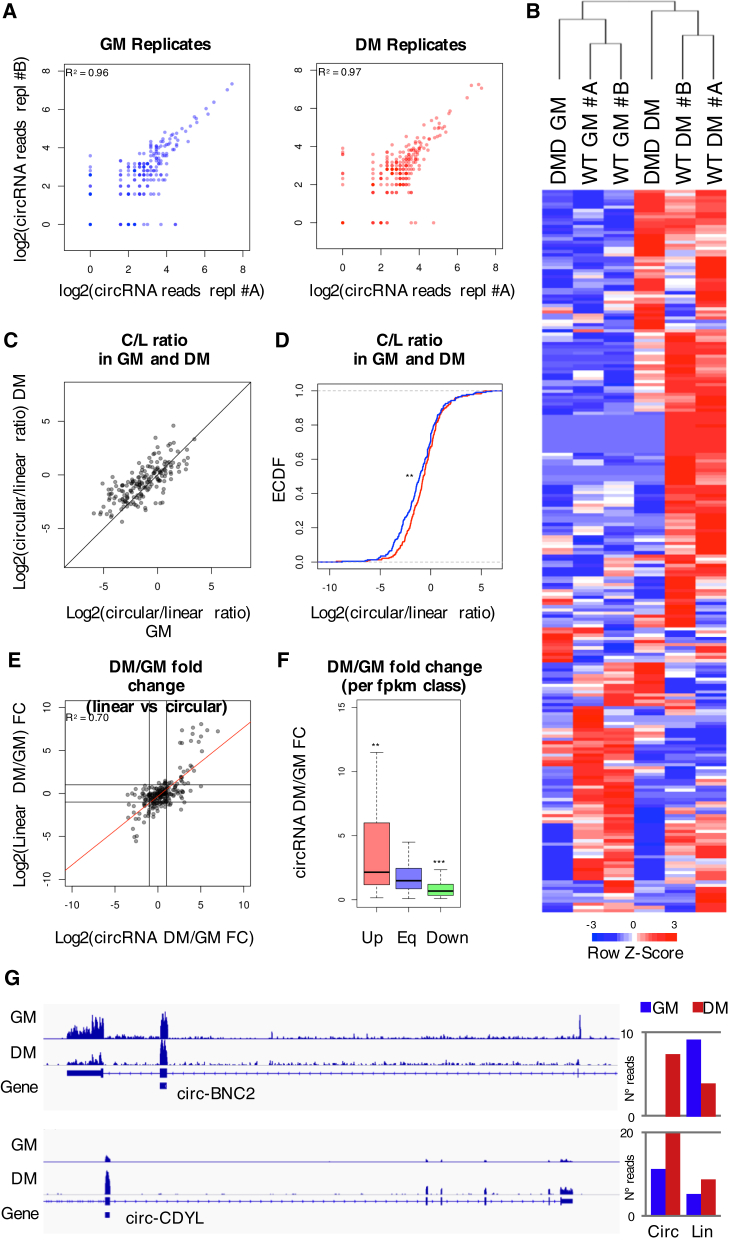
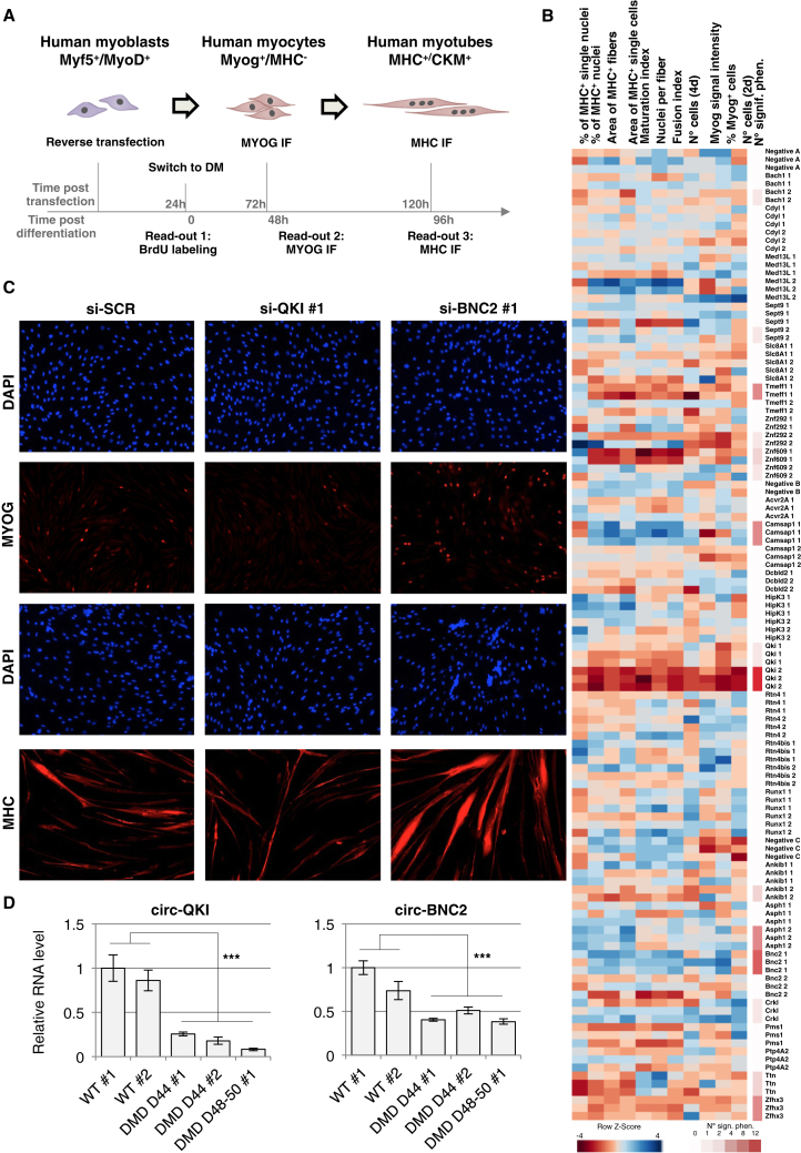
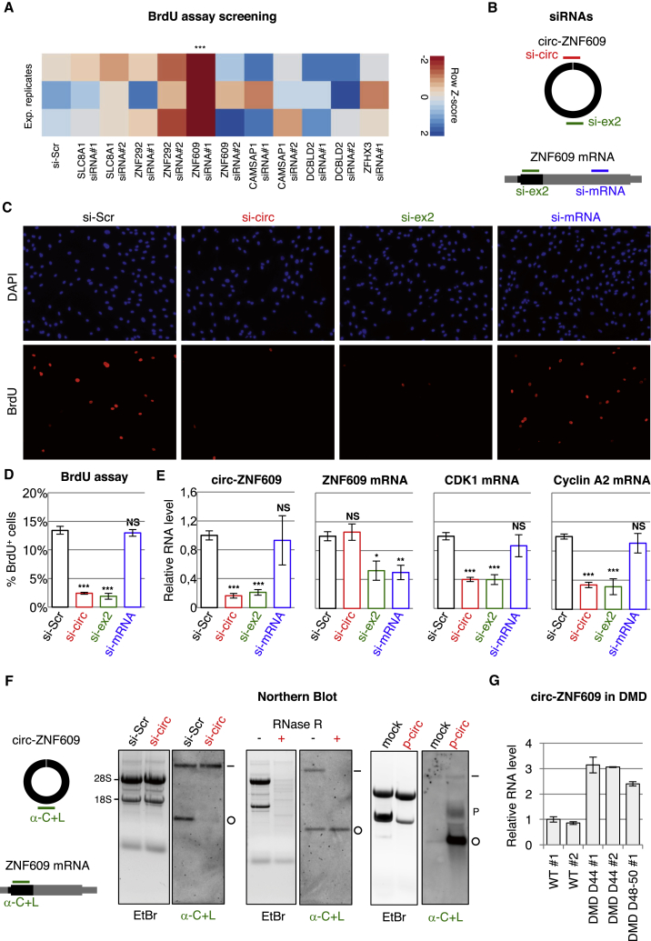
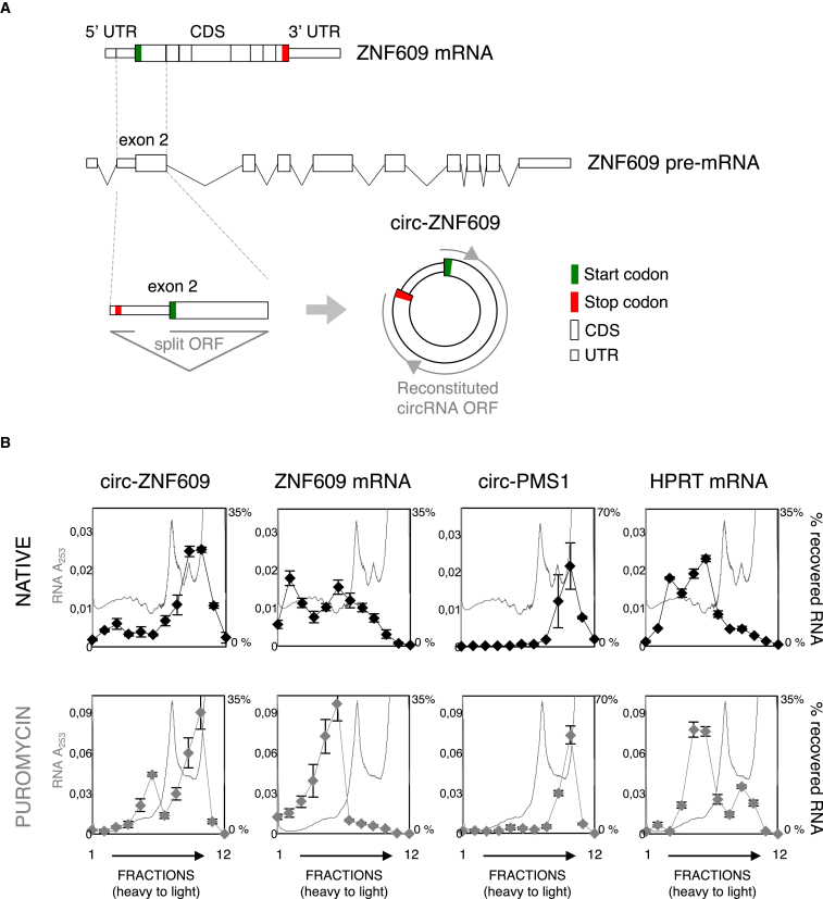
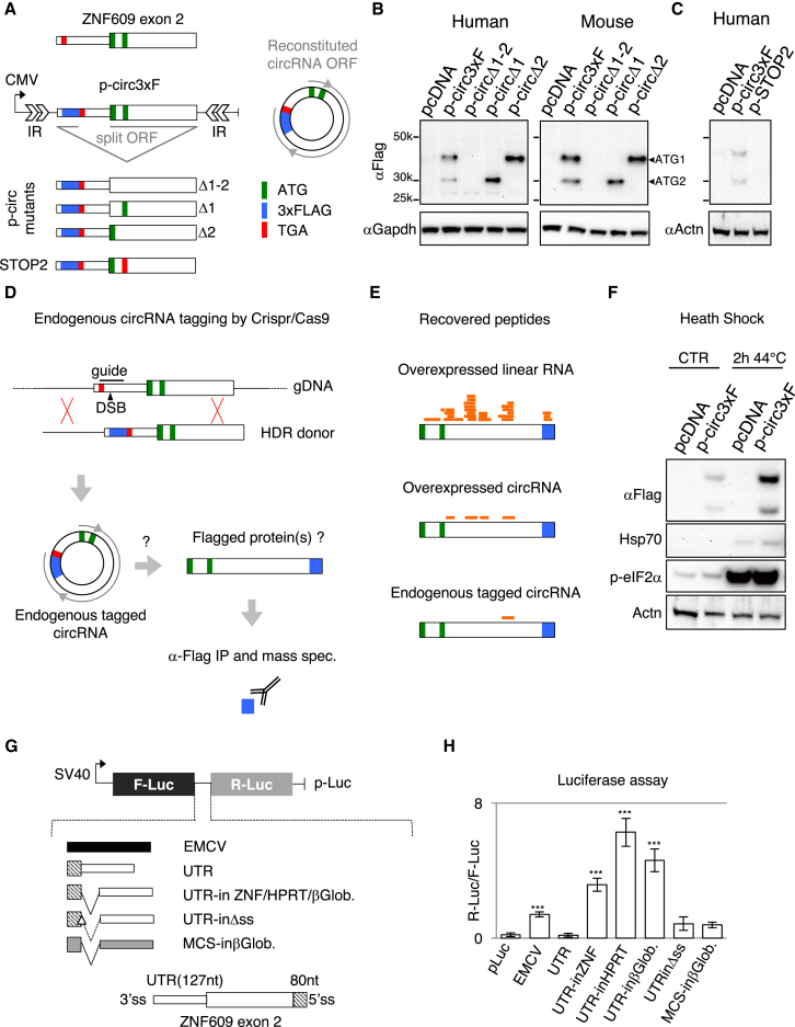
Circular RNAs (circRNAs) constitute a family of transcripts with unique structures and still largely unknown functions. Their biogenesis, which proceeds via a back-splicing reaction, is fairly well characterized, whereas their role in the modulation of physiologically relevant processes is still unclear. Here we performed expression profiling of circRNAs during in vitro differentiation of murine and human myoblasts, and we identified conserved species regulated in myogenesis and altered in Duchenne muscular dystrophy. A high-content functional genomic screen allowed the study of their functional role in muscle differentiation. One of them, circ-ZNF609, resulted in specifically controlling myoblast proliferation. Circ-ZNF609 contains an open reading frame spanning from the start codon, in common with the linear transcript, and terminating at an in-frame STOP codon, created upon circularization. Circ-ZNF609 is associated with heavy polysomes, and it is translated into a protein in a splicing-dependent and cap-independent manner, providing an example of a protein-coding circRNA in eukaryotes.

Keywords: DMD; cap independent; circRNA; circular RNA; muscle differentiation; myogenesis; non-coding RNA; proliferation; translation.


Figures

None
Graphical abstract
Figure 1
Figure 1
Identification of CircRNAs in Mammalian Myoblast Differentiation (A) Experimental and computational pipelines for the identification of circRNAs from human and mouse myoblasts (expressing MyoD and Myf5) and myotubes (expressing Myosin Heavy Chain [MHC] and Muscle Creatin Kinase [CKM]). Blue, rRNA and genomic reads; blue-red, reads across linear splice junctions; red, reads across back-splicing junctions. (B) Number of detected linear and circular splicing events per sample with bona fide circRNAs passing selection filters (see the STAR Methods). Samples are human primary and mouse C2C12 myoblasts (GM) and myotubes (DM) in two replicates (A and B). (C) Left panel: genomic annotation of circRNAs. Right panel: structural annotation of circRNAs mapping to coding regions is shown. (D) Overlap of unique circRNAs in human and mouse samples before and after (left and right) filtering for expression level higher than five unique reads in human samples. See also Figure S1 and Table S1.
Figure 2
Figure 2
Differential CircRNA Expression in Human Myoblast Differentiation and Disease (A) Scatterplots showing reads mapping to highly expressed circRNA junctions in two myoblast (GM, A and B, left) and myotube (DM, A and B, right) replicates. (B) Heatmap showing expression levels of selected circRNAs in WT and DMD myoblasts (GM, A and B) and myotubes (DM, A and B). Values are normalized as row Z scores. (C) Scatterplot showing circular/linear (C/L) ratio in GM versus DM, calculated as log2 of the number of reads mapping to a circular junction divided by the mean of reads mapping linearly at the same genomic coordinates. (D) Empirical cumulative distribution of the C/L ratio in GM (blue) and DM (red). (E) Scatterplot of fold change of reads mapping to circRNA junctions versus reads mapping to linear junctions at the same coordinates. Linear regression is shown in red. Black lines correspond to a fold change of 2. (F) Bar plot representing fold change of circRNA expression in myotubes versus myoblasts (DM/GM) grouped by differential expression of host gene according to Cuffdiff FPKMs. Two and three asterisks indicate a Wilcoxon-Mann-Whitney test-derived p value below 0.02 and 0.01, respectively. Up, upregulated genes; Eq, non-differentially expressed genes; down, downregulated genes (fold change > 2). (G) Left panels: coverage plots of genomic loci of circ-BNC2 and circ-CDYL. Right panels: bar plots show reads mapping to circular (Circ) and linear (Lin) splice sites for circ-BNC2 and circ-CDYL in GM (blue bars) and DM (red bars). See also Figure S2 and Table S1.
Figure 3
Figure 3
CircRNA Knockdown followed by Phenotypic Screening in Human Primary Myoblasts (A) Experimental workflow of the phenotypic screening strategy. (B) Heatmap showing selected phenotypes in color scale after Z score normalization. High values are shown in blue while low values are shown in red. Each column indicates one phenotype and each row indicates one sample. The number of detected significant phenotypes is shown in red scale on the right. (C) Human primary myoblasts were treated with either a control scramble siRNA (si-SCR) or with siRNAs against circ-QKI (si-QKI #1) and circ-BNC2 (si-BNC2 #1) and induced to differentiate for 48 (upper panels) and 96 hr (lower panels). A representative immunofluorescence for MYOG and MHC together with DAPI staining is shown. (D) RNA levels measured by qRT-PCR of circ-QKI and circ-BNC2 in human control (WT) and Duchenne (DMD) myocytes. RNA measurements were performed from biological triplicates of two WT (light gray bars) and three DMD primary cell lines (dark gray bars). See also Figure S3 and Table S3.
Figure 4
Figure 4
Circ-ZNF609 Controls Myoblast Proliferation (A) Human myoblasts were treated with either a control siRNA (si-Scr) or with siRNAs against the circRNA indicated; cells were maintained in growth conditions. The color scale represents the percentage of BrdU-positive cells measured in triplicate. (B) Schematic representation of the siRNAs used for circ-ZNF609 phenotype validation. The siRNA targeting specifically the circRNA is shown in red (si-circ), the siRNA targeting both the circRNA and the linear mRNA is shown in green (si-ex2), and the siRNA targeting only the mRNA is shown in blue (si-mRNA). (C) Human primary myoblasts were treated with either a control scramble siRNA (si-Scr) or the siRNAs shown in (B). A representative immunofluorescence for BrdU is shown in red and DAPI staining is shown in blue. (D) Percentage of BrdU incorporation levels in cells treated as in (C). (E) RNA quantification by qRT-PCR of circ-ZNF609, ZNF609 mRNA, and two markers of cell-cycle progression (CDK1 and Cyclin A2) after treatment with the siRNAs as in (C). (F) Left panel: schematic representation of the DIG-labeled probe used for northern blot analysis together with its location on the circular and linear forms of ZNF609 RNAs. Right panel: northern blots on 20 μg total RNA from primary myoblasts treated with either siRNAs (si-Scr and si-circ) or RNase R (– and +) are shown. The linear and the circular RNA forms are indicated aside the gels with the “–” and “o” symbols, respectively. As a control, northern blot was performed on 20 μg total RNA from myoblasts with the addition of 1 μg RNA from HeLa cells transfected with either an empty vector (mock) or a construct overexpressing circ-ZNF609 (p-circ). Each blot is shown in parallel to the EtBr staining of the agarose gel, where the migration of the 18S and 28S rRNAs is indicated. (G) RNA levels measured by qRT-PCR of circ-ZNF609 in human wild-type (WT) and Duchenne (DMD) myoblasts upon differentiation. RNA measurements were performed from biological duplicates of two WT and three DMD primary myoblasts. All values derive from three independent experiments unless differently stated. One asterisk indicates a Student’s t test-derived p value < 0.05, two asterisks indicate a p value < 0.02, and three asterisks a p value < 0.01. Bars show mean and SE. See also Figure S4.
Figure 5
Figure 5
Circ-ZNF609 RNA Is Associated with Polysomes (A) Schematic representation of the following: ZNF609 pre-mRNA (center), ZNF609 mRNA (above), and ZNF609 s exon (below) from which circ-ZNF609 (right) originates. The 5′ and 3′ UTRs are indicated by smaller boxes, while the coding region (CDS) is represented by larger boxes. Start and stop codons are shown in green and red, respectively. (B) Cytoplasmic extracts from proliferating human myoblasts, either untreated (native, black) or treated with puromycin (gray), were loaded on 15%–50% sucrose gradients. Absorbance at 253 nm was measured and fractions were collected. Fraction density decreases from left to right; the panels show one representative profile of three independent biological replicates. Individual fractions were analyzed by qRT-PCR and are represented as a percentage of total RNA in each fraction. Profiles are shown for circ-ZNF609 and ZNF609 mRNA. Circ-PMS1 and HPRT mRNA represent negative and positive controls, respectively. See also Figure S5.
Figure 6
Figure 6
Circ-ZNF609 Has Protein-Coding Activity (A) Schematic representation of the p-circ3xF construct. The second exon of the ZNF609 gene was cloned in a plasmid carrying inverted repeats (IRs) from the ZKSCAN1 gene (see the STAR Methods) in order to achieve circularization. A 3×FLAG tag was inserted upstream to the stop codon in order to obtain a flagged protein after circularization. Mutant derivatives with deleted ATGs are shown below. Start and stop codons are shown in green and red, respectively. A 3×FLAG-coding sequence is shown in blue. (B) Western blot analysis with an anti-FLAG antibody on proteins from HeLa (human) and N2a (mouse) cells transfected with p-circ3xF and its mutant derivatives. GAPDH was used as a loading control. (C) Western blot analysis with an anti-FLAG antibody on proteins from HeLa cells transfected with pcDNA, p-circ3xF, and its mutant derivative p-STOP2. ACTN was used as a loading control. (D) Schematic representation of the genome-editing approach for tagging the endogenous ZNF609 locus. Mouse embryonic stem cells were edited with homology-directed repair of a CRISPR/Cas9-induced double-strand break (DSB) with a DNA donor carrying a 3×FLAG-coding sequence (in blue) immediately upstream of the stop codon in exon 2. The edited cells were cultured and harvested for IP with an anti-FLAG antibody. The eluted proteins were run on SDS-PAGE (size selected between 30 and 40 kDa) and subjected to mass spectrometry. (E) Individual peptides detected by mass-spec are depicted in orange at their mapping position onto the protein sequence. Mass-spec data are shown for the overexpression of the linear ORF (see Figure S6A) by p-lin3xF (top), overexpression of the circular RNA from p-circ3xF (middle), and expression from the edited endogenous locus (bottom). (F) Representative western blot analysis with an anti-FLAG antibody on proteins from HeLa cells transfected with either pcDNA or p-circ3xF in normal (CTR) or heat shock conditions. Circ-ZNF609 RNA levels were the same in both conditions (data not shown). As controls of the heat shock response, blots were hybridized with Hsp70 and phosphorylated eIF2 alpha antibodies. Actinin was used as a loading control. (G) Schematic representation of the luciferase bicistronic constructs used. The SV40 promoter drives the transcription of Firefly (F-Luc) and Renilla (R-Luc) luciferase-coding regions in the control construct (p-Luc). The spacer of p-Luc was substituted with the IRES sequence of the Encephalomyocarditis virus (EMCV) or 80 nt from the 3′ portion of the second exon fused to the UTR of circ-ZNF609 (UTR). From UTR, additional constructs were raised carrying one intron from the ZNF609, HPRT, and β-globin (UTR-in ZNF/HPRT/βGlob.) genes. From UTR-in-βGlob. two other constructs were derived as follows: UTR-inΔss has a deletion of the 5′ splice site, while MCS-inβGlob. carries an unrelated sequence in place of the UTR. (H) Luciferase activity derived from cells transfected with the constructs described in (G). Values from three independent experiments are expressed as ratio of Renilla versus Firefly activities. Three asterisks represent a Student’s t test-derived p value < 0.01 between the value of each sample and the empty vector. Bars show mean and SE. See also Figure S6.

Comment in

  • RNA: Translated circular RNAs.
    Koch L. Koch L. Nat Rev Genet. 2017 May;18(5):272-273. doi: 10.1038/nrg.2017.27. Epub 2017 Apr 3. Nat Rev Genet. 2017. PMID: 28366936 No abstract available.

References

    1. Abe N., Matsumoto K., Nishihara M., Nakano Y., Shibata A., Maruyama H., Shuto S., Matsuda A., Yoshida M., Ito Y., Abe H. Rolling Circle Translation of Circular RNA in Living Human Cells. Sci. Rep. 2015;5:16435. - PMC - PubMed
    1. Ashwal-Fluss R., Meyer M., Pamudurti N.R., Ivanov A., Bartok O., Hanan M., Evantal N., Memczak S., Rajewsky N., Kadener S. circRNA biogenesis competes with pre-mRNA splicing. Mol. Cell. 2014;56:55–66. - PubMed
    1. Ballard D.H., Brown C.M. First Edition. Prentice Hall; 1982. Computer Vision.
    1. Bolger A.M., Lohse M., Usadel B. Trimmomatic: a flexible trimmer for Illumina sequence data. Bioinformatics. 2014;30:2114–2120. - PMC - PubMed
    1. Capel B., Swain A., Nicolis S., Hacker A., Walter M., Koopman P., Goodfellow P., Lovell-Badge R. Circular transcripts of the testis-determining gene Sry in adult mouse testis. Cell. 1993;73:1019–1030. - PubMed

MeSH terms