Review
. 2014 Jun 5;4(2):408-438.
doi: 10.3390/nano4020408.

Superparamagnetic Nanoparticles for Atherosclerosis Imaging

Affiliations
Review

Superparamagnetic Nanoparticles for Atherosclerosis Imaging

Fernando Herranz et al. Nanomaterials (Basel). .

Abstract

The production of magnetic nanoparticles of utmost quality for biomedical imaging requires several steps, from the synthesis of highly crystalline magnetic cores to the attachment of the different molecules on the surface. This last step probably plays the key role in the production of clinically useful nanomaterials. The attachment of the different biomolecules should be performed in a defined and controlled fashion, avoiding the random adsorption of the components that could lead to undesirable byproducts and ill-characterized surface composition. In this work, we review the process of creating new magnetic nanomaterials for imaging, particularly for the detection of atherosclerotic plaque, in vivo. Our focus will be in the different biofunctionalization techniques that we and several other groups have recently developed. Magnetic nanomaterial functionalization should be performed by chemoselective techniques. This approach will facilitate the application of these nanomaterials in the clinic, not as an exception, but as any other pharmacological compound.

Keywords: atherosclerosis plaque; cardiovascular imaging; chemoselective functionalization; iron oxide nanoparticles.


Conflict of interest statement

The authors declare no conflict of interest.

Figures

Figure 1
Figure 1
Steps in the synthesis of iron oxide nanoparticles (IONP) for preclinical atherosclerosis imaging. (A) Mixture of iron precursors and different surfactants in organic solvents; (B) Iron oxide nanoparticles in organic solvent; (C) Phase transfer to a water-based solution, in two steps or in a one-step phase transfer and functionalization (red arrow); (D) Functionalization of the nanoparticles for selective and/or multifunctional imaging; (E) Imaging of atherosclerotic plaque with iron oxide nanoparticles. EPR, enhanced permeability and retention.
Figure 2
Figure 2
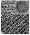
TEM images of 16-nm IONPs synthesized by the decomposition of organic precursors:  (A) a monolayer assembly; (B) a multilayer assembly; (C) High Resolution Transmission Electron Microscopy (HRTEM) image of a single Fe3O4 nanoparticle. Reproduced with permission from [13]. Copyright 2002, American Chemical Society.
Figure 3
Figure 3
Structure of common silane-based molecules for the ligand exchange approach.
Figure 4
Figure 4
Micelle approach for PAMAM-C12 coating of the oleic acid-capped iron oxide nanoparticles. Reproduced with permission from [142]. Copyright 2013, American Chemical Society.
Figure 5
Figure 5
Schematic structure and TEM imaging of HDL-iron oxide nanoparticles. Adapted with permission from [149]. Copyright 2008, American Chemical Society.
Figure 6
Figure 6
Direct chemical modification of the surfactant for oleic acid-coated IONPs, by (A) oxidation of the double bond and (B) olefin metathesis by the use of Hoveyda-Grubbs 2nd generation catalyst.
Figure 7
Figure 7
Synthesis of multifunctional IONPs by the direct chemical modification of oleic acid. The attachment of allergen Phl p5a and a fluorophore, via biotin-streptavidin interaction, was done by amide formation with the carboxylic groups generated. TEM image of the final IONPs (bottom left). Immunogenicity of the synthesized nanoparticles compared to non-functionalized particles, grass pollen extract and pure protein (bottom right). Reproduced from [87]. Copyright 2012, John Wiley & Sons, Ltd.
Figure 8
Figure 8
Evolution of atherosclerosis disease and the main targets at each step, according to the American Heart Assocciation (AHA). Reproduced with permission from [158]. Copyright 2008, Nature Publishing Group.
Figure 9
Figure 9
Fibrin-specific IONPs for the T1-weighted imaging of fibrin in atherosclerotic plaque. Reproduced with permission from [178]. Copyright 2009, American Chemical Society.

References

    1. Ittrich H., Peldschus K., Raabe N., Kaul M., Adam G. Superparamagnetic iron oxide nanoparticles in biomedicine: Applications and developments in diagnostics and therapy. RöFo Fortschritte Auf Dem Geb. Röntgenstrahlen Nukl. 2013;185:1149–1166. doi: 10.1055/s-0033-1335438. - DOI - PubMed
    1. Santhosh P.B., Ulrih N.P. Multifunctional superparamagnetic iron oxide nanoparticles: Promising tools in cancer theranostics. Cancer Lett. 2013;336:8–17. doi: 10.1016/j.canlet.2013.04.032. - DOI - PubMed
    1. Wang J., Huang Y., David A.E., Chertok B., Zhang L., Yu F., Yang V.C. Magnetic nanoparticles for MRI of brain tumors. Curr. Pharm. Biotechnol. 2012;13:2403–2416. doi: 10.2174/138920112803341824. - DOI - PubMed
    1. Kanwar R.K., Chaudhary R., Tsuzuki T., Kanwar J.R. Emerging engineered magnetic nanoparticulate probes for molecular MRI of atherosclerosis: How far have we come? Nanomedicine. 2012;7:899–916. doi: 10.2217/nnm.12.57. - DOI - PubMed
    1. Chauveau F., Cho T.H., Berthezène Y., Nighoghossian N., Wiart M. Imaging inflammation in stroke using magnetic resonance imaging. Int. J. Clin. Pharmacol. Ther. 2010;48:718–728. doi: 10.5414/CPP48718. - DOI - PubMed

LinkOut - more resources