. 2014 Oct 17;4(4):856-878.
doi: 10.3390/nano4040856.

Impact of the De-Alloying Kinetics and Alloy Microstructure on the Final Morphology of De-Alloyed Meso-Porous Metal Films

Affiliations

Impact of the De-Alloying Kinetics and Alloy Microstructure on the Final Morphology of De-Alloyed Meso-Porous Metal Films

Bao Lin et al. Nanomaterials (Basel). .

Abstract

Nano-textured porous metal materials present unique surface properties due to their enhanced surface energy with potential applications in sensing, molecular separation and catalysis. In this paper, commercial alloy foils, including brass (Cu85Zn15 and Cu70Zn30) and white gold (Au50Ag50) foils have been chemically de-alloyed to form nano-porous thin films. The impact of the initial alloy micro-structure and number of phases, as well as chemical de-alloying (DA) parameters, including etchant concentration, time and solution temperature on the final nano-porous thin film morphology and properties were investigated by electron microscopy (EM). Furthermore, the penetration depth of the pores across the alloys were evaluated through the preparation of cross sections by focus ion beam (FIB) milling. It is demonstrated that ordered pores ranging between 100 nm and 600 nm in diameter and 2-5 μm in depth can be successfully formed for the range of materials tested. The microstructure of the foils were obtained by electron back-scattered diffraction (EBSD) and linked to development of pits across the material thickness and surface during DA. The role of selective etching of both noble and sacrificial metal phases of the alloy were discussed in light of the competitive surface etching across the range of microstructures and materials tested.

Keywords: de-alloying (DA) kinetics; metal surface texture; micro-structure morphology relationship; through pores formation.


Conflict of interest statement

The authors declare no conflict of interest.

Figures
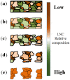
Figure 1
Figure 1
Schematic of the de-alloying (DA) process and pore propagation from the as a function of the grain distribution and composition: (a) pristine alloy microstructure with two alloy constituents distributed and mixed across a number of grains; (b) surface pitting and pore initiation; (c) DA of less noble atom rich exposed grains; (d) pore progression and through pore formation; and (e) final nano-porous materials. LNC: less noble content.
Figure 2
Figure 2
The scanning electron micrograph (SEM) images (in-plane view) of de-alloyed samples which de-alloyed with different etching solution (cross-section view has shown in Figures S2 and S3). 10 M HCl (pH = −1), 1 M HCl (pH = 0), 0.01 M HCl (pH = 2), deionized (DI) water (pH = 7), 1 × 10⁻⁵ M NaOH (pH = 9), 10 M NaOH (pH~14), 1 M NaOH (pH = 14) and 0.1 M NaOH (pH = 13). Scale bars all correspond to 500 nm.
Figure 3
Figure 3
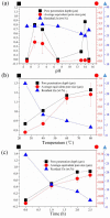
The impact of DA condition on surface pore size, penetrate depth and residual Zn content from energy dispersive spectroscopy (EDS): (a) pH value-based series de-alloyed foil de-alloyed with different etching solutions (10 M HCl (pH = −1), 1 M HCl (pH = 1), 0.01 M HCl (pH = 3), 10 M NaOH (pH = 15), 1 M NaOH (pH = 14) and 0.1 M NaOH (pH = 13)) for 2.5 h at 25 °C; (b) temperature-based series de-alloyed foil de-alloyed with 1 M NaOH for 2.5 h; and (c) time-based series de-alloyed foil de-alloyed with 1 M NaOH at 60 °C.
Figure 4
Figure 4
SEM image of de-alloyed CuZn15 and AuAg50: (a) DA CuZn15 with 1 M NaOH for 1 h at room temperature; (b) DA AuAg50 with 70% Nitric Acid for 10 min at room temperature; (c) cross-section view of (a); and (d) cross-section view of (b).
Figure 5
Figure 5
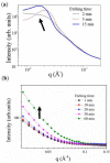
Small angle X-ray scattering (SAXS) analysis of in situ DA experiment on: (a) AuAg50; and (b) CuZn15.
Figure 6
Figure 6
X-ray diffraction (XRD) patterns of de-alloyed CuZn30 foil de-alloyed with 1 M NaOH and 1 M HCl: (a) 2θ = 35°–45°; (b) 2θ = 46°–53°; (c) 2θ = 62°–77°; and (d) 2θ = 81°–92°.
Figure 7
Figure 7
Schematic of the cross-flow contact rig: (a) non-recirculated flow; and (b) reference recirculated solution configurations.
Figure 8
Figure 8
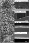
Surface and cross-section views of 1 M NaOH de-alloyed thin films with respective cross-flow surface velocities. Scale bars for the surface and cross section views correspond to 500 nm and 10 μm, respectively.
Figure 9
Figure 9
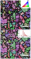
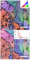
Electron back-scattered diffraction (EBSD) mapping on CuZn30 alloy: (a) before DA; and (b) after DA. Scale bars correspond to 5 μm. See Figure S10 for full scale image.
Figure 10
Figure 10
EBSD mapping of CuZn15: (a) before DA; and (b) after DA. Scale bars correspond to 5 μm. See Figure S11 for full scale image.

References

    1. Dumée L.F., He L., Lin B., Ailloux F.-M., Lemoine J.-B., Velleman L., She F., Duke M.C., Orbell J.D., Erskine G., et al. The fabrication and surface functionalization of porous metal frameworks—A review. J. Mater. Chem. A. 2013;1:15185–15206. doi: 10.1039/c3ta13240d. - DOI
    1. Kramer D., Viswanath R.N., Weissmüller J. Surface-stress induced macroscopic bending of nanoporous gold cantilevers. Nano Lett. 2004;4:793–796. doi: 10.1021/nl049927d. - DOI
    1. Biener J., Wittstock A., Zepeda-Ruiz L., Biener M., Zielasek V., Kramer D., Viswanath R., Weissmüller J., Bäumer M., Hamza A. Surface-chemistry-driven actuation in nanoporous gold. Nat. Mater. 2009;8:47–51. doi: 10.1038/nmat2335. - DOI - PubMed
    1. Jin H.-J., Weissmüller J. A material with electrically tunable strength and flow stress. Science. 2011;332:1179–1182. doi: 10.1126/science.1202190. - DOI - PubMed
    1. Newman R., Mehta A. An AC impedance study of the de-alloying of Fe-Ni alloys, and its relevance to chloride SCC of stainless steels. Corros. Sci. 1988;28:1183–1187. doi: 10.1016/0010-938X(88)90127-8. - DOI