. 2015 Dec 25;6(1):2.
doi: 10.3390/nano6010002.

Effects of Particle Hydrophobicity, Surface Charge, Media pH Value and Complexation with Human Serum Albumin on Drug Release Behavior of Mitoxantrone-Loaded Pullulan Nanoparticles

Affiliations

Effects of Particle Hydrophobicity, Surface Charge, Media pH Value and Complexation with Human Serum Albumin on Drug Release Behavior of Mitoxantrone-Loaded Pullulan Nanoparticles

Xiaojun Tao et al. Nanomaterials (Basel). .

Abstract

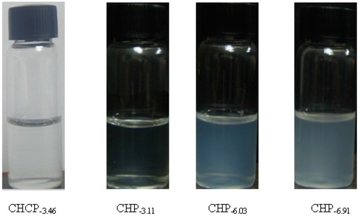
We prepared two types of cholesterol hydrophobically modified pullulan nanoparticles (CHP) and carboxyethyl hydrophobically modified pullulan nanoparticles (CHCP) substituted with various degrees of cholesterol, including 3.11, 6.03, 6.91 and 3.46 per polymer, and named CHP-3.11, CHP-6.03, CHP-6.91 and CHCP-3.46. Dynamic laser light scattering (DLS) showed that the pullulan nanoparticles were 80-120 nm depending on the degree of cholesterol substitution. The mean size of CHCP nanoparticles was about 160 nm, with zeta potential -19.9 mV, larger than CHP because of the carboxyethyl group. A greater degree of cholesterol substitution conferred greater nanoparticle hydrophobicity. Drug-loading efficiency depended on nanoparticle hydrophobicity, that is, nanoparticles with the greatest degree of cholesterol substitution (6.91) showed the most drug encapsulation efficiency (90.2%). The amount of drug loading increased and that of drug release decreased with enhanced nanoparticle hydrophobicity. Nanoparticle surface-negative charge disturbed the amount of drug loading and drug release, for an opposite effect relative to nanoparticle hydrophobicity. The drug release in pullulan nanoparticles was higher pH 4.0 than pH 6.8 media. However, the changed drug release amount was not larger for negative-surface nanoparticles than CHP nanoparticles in the acid release media. Drug release of pullulan nanoparticles was further slowed with human serum albumin complexation and was little affected by nanoparticle hydrophobicity and surface negative charge.

Keywords: HSA; degree of substitution; pullulan nanoparticles; surface charge.


Conflict of interest statement

The authors declare no conflict of interest.

Figures

Figure 1
Figure 1
Chemical structure of pullulan.
Figure 2
Figure 2
Structural graph of pullulan nanoparticle.
Figure 3
Figure 3
Sketch illustration of human serum albumin (HSA) complexation with pullulan nanoparticles.
Figure 4
Figure 4
The optical appearance of pullulan nanoparticles with different hydrophobicity and surface charge.
Figure 5
Figure 5
Transmission electron microscopy (TEM) of CHP−6.91 (a); CHP−6.03 (b); CHP−3.11 (c) and CHCP (d) nanoparticles.
Figure 6
Figure 6
Size distribution of CHP−6.91 (a); CHP−6.03 (b); CHP−3.11 (c) and CHCP (d) nanoparticles.
Figure 7
Figure 7
Zeta potential of CHP−6.91 (a); CHP−6.03 (b); CHP−3.11 (c) and CHCP (d) nanoparticles.
Figure 8
Figure 8
TEM of CHP−6.03 nanoparticles loading mitoxantrone.
Figure 9
Figure 9
The mitoxantrone (MTO) release of pullulan nanoparticles in phosphate buffered saline (PBS) at 37 °C in vitro (: free mitoxantrone, : CHP−3.11, : CHCP, : CHP−6.03, : CHP−6.91).
Figure 10
Figure 10
The mitoxantrone (MTO) release from pullulan nanoparticles in PBS buffer (pH 6.8) at 37 °C in vitro and acetate buffer (pH 4.0) (: CHP−3.11, : CHCP; pH 6.8), (: CHCP, : CHP−3.11; pH 4.0).
Figure 11
Figure 11
The mitoxantrone (MTO) release of pullulan nanoparticles upon human serum albumin (HSA) complexation in PBS at 37 °C in vitro (: CHP−6.91, : CHP−3.11, : CHCP); and in the HSA release media (: CHP−6.91, : CHP−3.11, : CHCP).
Figure 12
Figure 12
Drug released from nanoparticles upon HSA complexation and in the HSA release media.

Similar articles

Cited by

References

    1. Yang T., Choi M., Cui F., Kim J.S., Chung S.J., Shim C.K., Kim D.D. Preparation and evaluation of paclitaxel-loaded PEGylated immunoliposome. J. Control. Release. 2007;120:169–177. doi: 10.1016/j.jconrel.2007.05.011. - DOI - PubMed
    1. Koneru B., Shi Y., Wang Y.C., Chavala S.H., Miller M.L., Holbert B., Conson M., Ni A., Pasqua A.J.D. Tetracycline-Containing MCM-41 Mesoporous Silica Nanoparticles for the Treatment of Escherichia coli. Molecules. 2015;20:19690–19698. doi: 10.3390/molecules201119650. - DOI - PMC - PubMed
    1. Bennet D., Marimuthu M., Kim S., An J. Dual drug-loaded nanoparticles on self-integrated scaffold for controlled delivery. Int. J. Nanomed. 2012;7:3399–3419. - PMC - PubMed
    1. Bhirde A.A., Kapoor A., Liu G., Iglesias-Bartolome R., Jin A., Zhang G.F., Xing R.J., Lee S., Leapman R.D., Gutkind S.J., Chen X.Y. Nuclear mapping of nano-drug delivery systems in dynamic cellular environments. ACS Nano. 2012;6:4966–4972. doi: 10.1021/nn300516g. - DOI - PMC - PubMed
    1. Ptail Y., Sadhukha T., Ma L., Panyam J. Nanoparticle-mediated simultaneous and targeted delivery of paclitaxel and tarquidar overcomes tumor drug resistance. J. Control. Release. 2009;136:21–29. doi: 10.1016/j.jconrel.2009.01.021. - DOI - PubMed

LinkOut - more resources