. 2016 Jan 5;6(1):8.
doi: 10.3390/nano6010008.

Targeted Sterically Stabilized Phospholipid siRNA Nanomedicine for Hepatic and Renal Fibrosis

Affiliations

Targeted Sterically Stabilized Phospholipid siRNA Nanomedicine for Hepatic and Renal Fibrosis

Fatima Khaja et al. Nanomaterials (Basel). .

Abstract

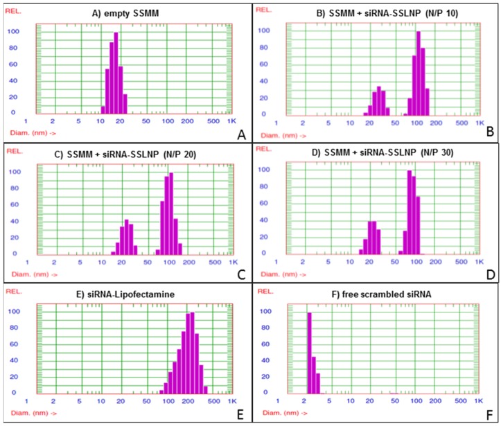
Since its discovery, small interfering RNA (siRNA) has been considered a potent tool for modulating gene expression. It has the ability to specifically target proteins via selective degradation of messenger RNA (mRNA) not easily accessed by conventional drugs. Hence, RNA interference (RNAi) therapeutics have great potential in the treatment of many diseases caused by faulty protein expression such as fibrosis and cancer. However, for clinical application siRNA faces a number of obstacles, such as poor in vivo stability, and off-target effects. Here we developed a unique targeted nanomedicine to tackle current siRNA delivery issues by formulating a biocompatible, biodegradable and relatively inexpensive nanocarrier of sterically stabilized phospholipid nanoparticles (SSLNPs). This nanocarrier is capable of incorporating siRNA in its core through self-association with a novel cationic lipid composed of naturally occuring phospholipids and amino acids. This overall assembly protects and delivers sufficient amounts of siRNA to knockdown over-expressed protein in target cells. The siRNA used in this study, targets connective tissue growth factor (CTGF), an important regulator of fibrosis in both hepatic and renal cells. Furthermore, asialoglycoprotein receptors are targeted by attaching the galactosamine ligand to the nanocarries which enhances the uptake of nanoparticles by hepatocytes and renal tubular epithelial cells, the major producers of CTGF in fibrosis. On animals this innovative nanoconstruct, small interfering RNA in sterically stabilized phospholipid nanoparticles (siRNA-SSLNP), showed favorable pharmacokinetic properties and accumulated mostly in hepatic and renal tissues making siRNA-SSLNP a suitable system for targeting liver and kidney fibrotic diseases.

Keywords: fibrosis; galactosamine; hepatic stellate cells; siRNA; sterically stabilized phospholipid nanoparticles.


Conflict of interest statement

The authors declare no conflict of interest.

Figures

Figure 1
Figure 1
small interfering RNA in sterically stabilized phospholipid nanoparticles (siRNA-SSLNP) optimization in vitro: Representative particle size distribution of (A) Unimodal size distribution of empty SSMM (sterically stabilized mixed micelles); (B) Empty SSMM and siRNA-SSLNP with N/P ratio of 10; (C) Empty SSMM and siRNA-SSLNP with N/P ratio of 20; (D) Empty SSMM and siRNA-SSLNP with N/P ratio of 30; (E) siRNA lipofectamine (siRNA-Lipofectamine) complex; (F) Free siRNA molecules.
Figure 2
Figure 2
siRNA-SSLNP in vitro characterization: (A) Gel retardation assay of different formulations of siRNA, containing 200 nM siRNA per sample, on TBE-urea 15% gel , at a voltage of 180 V for 60 min, then stained with 1:500 SYBR Green-II in TBE with mild agitation for 30 min; (B) Fluorescence intensities measured by SYBR Green-II exclusion assay of SSMM and siRNA-SSLNP complexes at varying N/P ratios and siRNA with lipofectamine (LF) showing percent of un-incorporated siRNA (* p < 0.05 vs. free siRNA; mean ± SD; n = 3 replicates/group); (C) Gel retardation assay of different siRNA formulations after treatment with RNase; (D) Fluorescence intensities measured by SYBR Green-II fluorescence assay of different siRNA formulations after treatment with RNase (* p < 0.05 vs. free siRNA; † p < 0.05 vs. siRNA-lipofectamine; mean ± SD; n = 3 replicates/group).
Figure 3
Figure 3
Physicochemical characterization of siRNA-SSLNP-GalN: (A) Particle size distribution showing SSLNP-GalN peak at 91 ± 13 nm; (B) Transmission electron microscopy (TEM) image of siRNA-SSLNP-GalN, scale bar = 100 nm; (C) Results of SYBR Green-II exclusion assay. Bars represent percentage of siRNA before (un-incorporated) and after treatment with RNase enzyme (mean ± SD; n = 3 replicates/group).
Figure 4
Figure 4
Cell uptake and cytotoxicity assays: (A) Hepatic Hep-G2 cell uptake of FAM-labeled siRNA in various complexes. Changes in FACS histogram indicative of siRNA positive cells (upper), bars represent quantitative analysis of FACS histogram as a percentage siRNA-positive cells (lower). (* p < 0.05 vs. free siRNA and untreated control, † p > 0.05 meaning no statistical significance vs. siRNA-LF treated cells); (B) Cytotoxicity of siRNA in various complexes against primary hepatic stellate cells HSC at different siRNA concentration as determined by membrane integrity (LDH) assay; (C) Relative Hep-G2 cell viability expressed as a percentage of untreated control as a measure of cytotoxicity of siRNA complexes using MTS assay after 72 h incubation; (D) Cytotoxicity of different siRNA formulations after incubation with renal HK-2 cells for 72 h; (E) Cell proliferation kinetics of HK-2 cells after treatment with different formulations at siRNA concentration equivalent to 250 nM assessed at 24, 48, and 72 h time points (* p < 0.05 vs. siRNA-LF, data on B–D presented as mean ± SD; n = 3 replicates/group).
Figure 5
Figure 5
Protein downregulation: (A) Reduction of connective tissue growth factor (CTGF) expression in human hepatic Hep-G2 cells 24 h post transfection with anti CTGF-siRNA in different complexes; (B) Reduction of extracellular matrix (ECM) collagen expression in primary human hepatic stellate cells (HSC) 24 h post transfection with anti CTGF-siRNA in different complexes; (C) GTGF protein downregulation in renal tubular HK-2 cells, activated with transforming growth factor β1 (TGF-β1), 72 h post transfection with anti CTGF-siRNA in different complexes; (D) Reduction in ECM collagen expression by TGFβ activated HK-2 cells 72 h post transfection with anti CTFG-siRNA in different complexes. (Data are expressed as percent of the untreated control; mean ± SD; n = 3/treatment; * p < 0.05 vs. free siRNA at a corresponding siRNA dose; # p > 0.05 or statistically not significant vs. siRNA-lipofectamine (LF); ns—non significant among the groups indicated). The scrambled siRNA treatment showed no significance as compared to untreated controls and therefore control refers to untreated controls (Data not plotted).
Figure 6
Figure 6
Reversal of primary hepatic stellate cell (HSC) activation: (A) Down-regulation of collagen I; (B) Collagen III; and (C) α-smooth muscle actin (α-SMA) protein expression indicative of the reversal of activated myofibroblasts to quiescent stellate cells. Activated HSC were transfected with connective tissue growth factor (CTGF-siRNA) in various formulations. Standard immunocytochemistry performed 24 h post-transfection, cells were probed with primary antibodies followed by secondary Alexa-Fluor 488 (green) labeled antibody, then DAPI for nuclear staining (blue).
Figure 7
Figure 7
Biodistribution of different siRNA formulations compared to free Cy5 fluorophore over 24 h periods in (A) liver; (B) lung; (C) spleen; (D) heart; and (E) kidneys. Targeted formulation (siRNA-SSLNP-GalN) shows significant concentrations in liver and kidneys over observation period (n = 4 for each time point; * p < 0.05 vs. free siRNA treated animals, † p < 0.05 vs. free Cy5 treated animals); (F) Plasma concentration vs. time after single intravenous administration of various Cy-5 labeled formulations in Balb/c mice. (Data are presented as mean ± SD; n = 4 animals/each time point, MFI-Mean fluorescence intensity).

References

    1. Gavrilov K., Saltzman W.M. Therapeutic siRNA: Principles, challenges, and strategies. Yale J. Biol. Med. 2012;85:187–200. - PMC - PubMed
    1. Lam J.K., Chow M.Y., Zhang Y., Leung S.W. siRNA Versus miRNA as Therapeutics for Gene Silencing. Mol. Ther. Nucleic Acids. 2015;4 doi: 10.1038/mtna.2015.23. - DOI - PMC - PubMed
    1. Ozpolat B., Sood A., Lopez-Berestein G. Nanomedicine based approaches for the delivery of siRNA in cancer. J. Intern. Med. 2010;267:44–53. doi: 10.1111/j.1365-2796.2009.02191.x. - DOI - PubMed
    1. Ameyar-Zazoua M., Guasconi V., Ait-Si-Ali S. siRNA as a route to new cancer therapies. Expert Opin. Biol. Ther. 2005;5:221–224. doi: 10.1517/14712598.5.2.221. - DOI - PubMed
    1. Lin Q., Chen J., Zhang Z., Zheng G. Lipid-based nanoparticles in the systemic delivery of siRNA. Nanomedicine. 2014;9:105–120. doi: 10.2217/nnm.13.192. - DOI - PubMed

LinkOut - more resources