Review
. 2017 Mar;33(2):107-118.
doi: 10.6515/acs20160818a.

Three-Dimensional Echocardiography: Current Status and Real-Life Applications

Affiliations
Review

Three-Dimensional Echocardiography: Current Status and Real-Life Applications

Victor Chien-Chia Wu et al. Acta Cardiol Sin. 2017 Mar.

Abstract

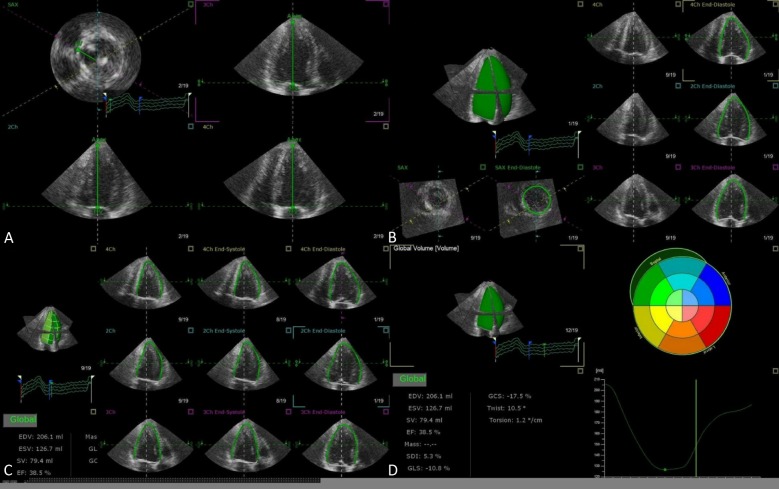
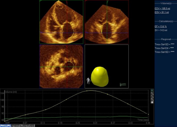
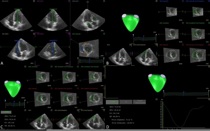
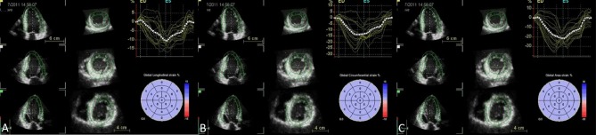
The use of cardiac ultrasound is fundamental to the understanding of normal heart function and crucial to pathophysiological diagnosis. The growing availability of 3D echocardiography (3DE) over the last decade has allowed its applications to expand from establishing reference values for chamber size and elucidating ventricular mechanics, to assessing valvular disease severity and playing pivotal roles in interventional procedures. Several important advantages of 3DE include eliminating geometric assumptions, quantifying complex geometric shape volumes, viewing structures from any perspective, assessing lesion in simultaneous multiplanes or multislice mode, all of which are not possible with traditional 2D echocardiography (2DE). Real-time 3DE has been shown to be simple, accurate, reproducible, and versatile, and generally has superior outcome prognosis compared to the 2DE.

Keywords: 3D echocardiography; Chamber mechanics; Valvular lesions.


Figures

Figure 1
Figure 1
Representative case of LV quantification step-by-step using TomTec 4D LV Analysis software. View alignment (A), Beutel revision (B), Tracking revision (C), Analysis (D). LV, left ventricular.
Figure 2
Figure 2
Representative case of LA quantification using Philips QLab. From the full-volume datasets, 2 orthogonal long-axis and 1 short-axis views of the LA at end-diastole and end-systole were selected. The software automatically determined the LA wall in 3D space using the deformable shell model and made time domain LA volume curve, from which maximal and minimal LA volumes. Manual adjustment when inadequate tracking of the LA wall was observed. LA, left atrial.
Figure 3
Figure 3
Representative case of RV quantification step-by-step using TomTec 4D RV Analysis software. View adjustment (A), Beutel revision (B), Tracking revision (C), Analysis (D). RV, right ventricular.
Figure 4
Figure 4
After region of interests are marked manually, software automatically traced on the endocardial border. Manual adjustments are required when software border detection are suboptimal. Next, epicardial border are manually determined for region of interest. The computer then analyzes 3D speckle-tracking in frame-by-frame analysis with a final 17-segment bull’s eye map of strain values displayed. Longitudinal strain (A), circumferential strain (B), and area strain (C).
Figure 5
Figure 5
Mitral valve regurgitation visualized as vena contracta width (VCW) along the long axis of mitral regurgitation jet in anterior-posterior (AP) and commissure-commissure (CC) views and as vena contracta area (VCA) along short-axis (SAX) view in transesophageal 3DE. The VCA is seen not circular but ellipsoidal inen face SAX view.
Figure 6
Figure 6
Assessment of aortic root by transesophageal 3D echocardiography. As seen in the last row of panel (D), the en face cross-sectional view of aortic annulus is oval rather than circular.
Figure 7
Figure 7
Simultaneous display of multiplane by 3D echocardiography. Top row, from left: parasternal long-axis, basal short-axis, mid short-axis views. Bottom row, from left: apical 4-chamber, apical 2-chamber, and apical 3-chamber views.
Figure 8
Figure 8
Simultaneous display of multi-slice by 3D echocardiography. Nine-equidistant 2D short-axis images from LV apex (top left) to base (bottom right).

References

    1. von Ramm OT, Smith SW. Real time volumetric ultrasound imaging system. J Digital Imaging. 1990;3:261–266. - PubMed
    1. Lang RM, Mor-Avi V, Dent JM, Kramer CM. Three-dimensional echocardiography: is it ready for everyday clinical use? JACC Cardiovasc Imaging. 2009;2:114–117. - PubMed
    1. Mor-Avi V, Sugeng L, Lang RM. Real-time 3-dimensional echocardiography: an integral component of the routine echocardiographic examination in adult patients? Circulation. 2009;119:314–329. - PubMed
    1. Lang RM, Badano LP, Tsang W, et al. American Society of Echocardiography; European Association of Echocardiography. EAE/ASE recommendations for image acquisition and display using three-dimensional echocardiography. JASE. 2012;25:3–46. - PubMed
    1. Jenkins C, Bricknell K, Hanekom L, Marwick TH. Reproducibility and accuracy of echocardiographic measurements of left ventricular parameters using real-time three-dimensional echocardiography. J Am Coll Cardiol. 2004;44:878–886. - PubMed

LinkOut - more resources