

Capitate Chondroblastoma: A Case Report and Review of the Literature
- PMID: 28344536
- PMCID: PMC5349402
- DOI: 10.1177/1558944716642762
Capitate Chondroblastoma: A Case Report and Review of the Literature
Abstract
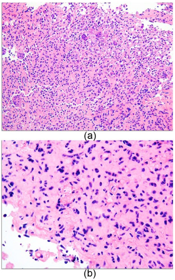
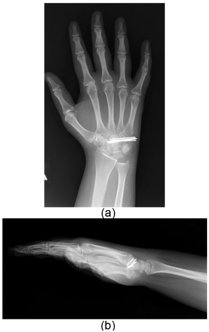
Background: Chondroblastomas are benign tumors that typically occur in the epiphysis of long bones. Carpal bone chondroblastomas are very rare and are known to have less aggressive behavior with no evidence of recurrence reported. Methods: We present a case of a recurrent chondroblastoma in the capitate that was treated with repeat curettage, application of phenol, and bone grafting. Results: At 3 years post surgery, the patient is disease free with excellent functional return. Conclusion: Chondroblastomas are rare within the carpus. We present a review of the literature detailing their occurrence and treatment.
Keywords: arthrodesis; capitate chondroblastoma; recurrence.
Conflict of interest statement
Declaration of Conflicting Interests: The authors declared no potential conflicts of interest with respect to the research, authorship, and/or publication of this article.
Figures


References
-
- Dahlin DC, Ivins JC. Benign chondroblastoma. A study of 125 cases. Cancer. 1972;30:401-413. - PubMed
-
- Daly KE, Chow JW, Vickers RH. Excision of the hamate for an unusual hand tumour. J Hand Surg Br. 1993;18:606-608. - PubMed
-
- Davila JA, Amrami KK, Sundaram M, Adkins MC, Unni KK. Chondroblastoma of the hands and feet. Skeletal Radiol. 2004;33:582-587. - PubMed
-
- de Silva MV, Reid R. Chondroblastoma: varied histologic appearance, potential diagnostic pitfalls, and clinicopathologic features associated with local recurrence. Ann Diagn Pathol. 2003;7:205-213. - PubMed
-
- Oishi SN, Muzaffar AR, Carter PR. Treatment of Kienbock’s disease with capitohamate arthrodesis: pain relief with minimal morbidity. Plast Reconstr Surg. 2002;109:1293-1300. - PubMed
Publication types
MeSH terms
LinkOut - more resources
Full Text Sources
Other Literature Sources
Medical
