. 2017 Mar 23:6:2.
doi: 10.1186/s13630-017-0048-6. eCollection 2017.

Abnormal glycosylation in Joubert syndrome type 10

Affiliations

Abnormal glycosylation in Joubert syndrome type 10

Megan S Kane et al. Cilia. .

Abstract

Background: The discovery of disease pathogenesis requires systematic agnostic screening of multiple homeostatic processes that may become deregulated. We illustrate this principle in the evaluation and diagnosis of a 5-year-old boy with Joubert syndrome type 10 (JBTS10). He carried the OFD1 mutation p.Gln886Lysfs*2 (NM_003611.2: c.2656del) and manifested features of Joubert syndrome.

Methods: We integrated exome sequencing, MALDI-TOF mass spectrometry analyses of plasma and cultured dermal fibroblasts glycomes, and full clinical evaluation of the proband. Analyses of cilia formation and lectin staining were performed by immunofluorescence. Measurement of cellular nucleotide sugar levels was performed with high-performance anion-exchange chromatography with pulsed amperometric detection. Statistical analyses utilized the Student's and Fisher's exact t tests.

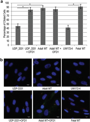
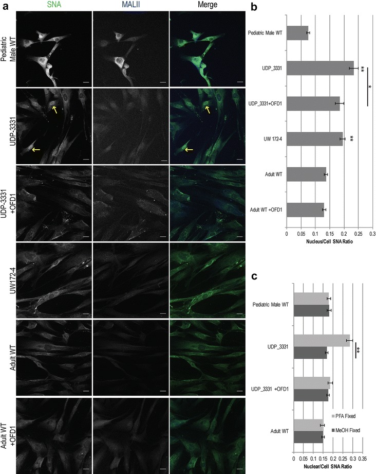
Results: Glycome analyses of plasma and cultured dermal fibroblasts identified abnormal N- and O-linked glycosylation profiles. These findings replicated in two unrelated males with OFD1 mutations. Cultured fibroblasts from affected individuals had a defect in ciliogenesis. The proband's fibroblasts also had an abnormally elevated nuclear sialylation signature and increased total cellular levels of CMP-sialic acid. Ciliogenesis and each glycosylation anomaly were rescued by expression of wild-type OFD1.

Conclusions: The rescue of ciliogenesis and glycosylation upon reintroduction of WT OFD1 suggests that both contribute to the pathogenesis of JBTS10.

Keywords: CMP-sialic acid; Ciliopathy; Glycosylation; Joubert syndrome; Molar tooth sign.


Figures

Fig. 1
Fig. 1
Clinical and molecular evidence for Joubert syndrome type 10 diagnosis. a Magnetic resonance imaging of the proband, UDP-3331, reveals the hallmark “molar tooth sign” of the superior cerebellar peduncles (arrow). b Sanger sequencing of both the proband, UDP-3331, and his mother, UDP-4786, confirms an OFD1 single-nucleotide deletion (c.2656del) in the proband and maternal inheritance. c Analysis of mRNA transcripts from adult control cells (“WT”), UDP-3331, and UDP-3331 + OFD1 rescue shows reduced mRNA levels in the proband. Error bars represent standard error of four replicates; data are normalized to GAPDH expression and plotted relative to WT OFD1 expression levels. Asterisk indicates p < 0.001 for two-tailed, heteroscedastic Student’s t test. d Western blot analysis of OFD1 levels in UDP-3331, UDP-3331 + OFD1 and adult WT primary fibroblast lines; one representative blot with OFD1 and α-tubulin is shown in the top panels; average of 3 replicate blots with independent lysates is shown in the lower panel. OFD1 levels are normalized to α-tubulin and plotted relative to WT OFD1 protein levels
Fig. 2
Fig. 2
Cellular morphology and analysis of JBTS10 skin fibroblasts. a Co-localization analysis of OFD1 (magenta) signals in proband, proband rescued, and adult WT cells with ɣ-tubulin (red) and PCM1 (green). Inset higher magnification of the region outlined with dotted lines. b Immunofluorescence staining for OFD1 (green) and α-tubulin (red) in fibroblasts of the proband (UDP-3331), proband rescued (UDP-3331 + OFD1), and adult WT control. c Comparison of endoplasmic reticulum structure (KDEL, green) in UDP-3331 and WT cells. d Comparison of the cis- (GM-130, green) and trans- (TGN-46, red) Golgi in UDP-3331 and WT cells. DNA was stained with Hoechst 33,342 (blue). All scale bars represent 20 µm
Fig. 3
Fig. 3
Analysis of ciliogenesis in JBTS10 and control skin fibroblasts. a The percentage of ciliated cells following 48 h of serum starvation in UDP-3331, UDP-3331 + OFD1 rescue, adult WT, adult WT + OFD1 overexpression, UW172-4, and fetal WT control cells. Error bars represent the 95% confidence interval for the average of three independent experiments. Asterisks indicate p < 0.001, Student’s t test. b Representative images of ciliated cells for the indicated cell lines. Acetylated tubulin (red) and ɣ-tubulin (green) mark the cilia and basal body, respectively. DNA was stained with DAPI (blue). Scale bars 10 µm
Fig. 4
Fig. 4
Lectin staining for sialic acid epitopes in JBTS10 and control skin fibroblasts. a Lectin staining of α-2,6-sialic acid (SNA, green) and α-2,3-sialic acid (MALII, blue) linkages in affected, rescued and control cells, as indicated. Arrows indicate atypical nuclear staining by SNA in UDP-3331. Scale bars 20 µm. b Quantification of the fluorescence signal for SNA in nucleus relative to the entire cell for affected, rescued, and control cells under paraformaldehyde fixation conditions. Error bars represent standard error. Single asterisks indicate p < 0.05; double asterisks indicate p < 0.001 relative to both adult and pediatric male controls (unpaired, heteroscedastic t test). c Comparison of methanol and paraformaldehyde fixation on the relative SNA staining intensity of the nucleus vs. the whole cell. Double asterisks indicate p < 0.001(unpaired, heteroscedastic t test). Error bars represent standard error
Fig. 5
Fig. 5
Nucleotide sugar analysis in JBTS10 and control skin fibroblasts. a Representative HPAEC trace for UDP-3331, UDP-3331 + OFD1 rescue, adult WT, adult WT + OFD1 overexpression, pediatric male control cells, and 500 nM standard CMP-sialic acid. b Quantification of CMP-sialic acid levels in affected and control cells (in μM) (single asterisks indicate p < 0.005 in an unpaired, heteroscedastic t test). Error bars represent standard error
Fig. 6
Fig. 6
Neuraminidase treatment of adult WT primary cells. a Average cilia length of WT cells with and without neuraminidase (±Neuase) treatment to remove sialic acids from glycan chains; neuraminidase treatment significantly reduced average cilia length by ~1 µm. Error bars represent standard error; p < 0.001 as determined in an unpaired, heteroscedastic t test. b The percentage of ciliated cells is decreased after neuraminidase treatment; p < 0.05 as determined by a two-tailed Fisher’s Exact t test; error bars represent the 95% confidence interval for the binomial distribution represent by the sample population. c, d Representative images of WT fibroblasts without (c) and with (d) neuraminidase treatment. Cilia are stained with ARL13B antibody (red) and ɣ-tubulin (green). Scale bars 20 µm. e Lectin staining for sialic acid with SNA (green) and MAL II (white) and galactose with PNA (red) in WT fibroblasts with DRAQ5 DNA counterstaining (blue). f Representative image of SNA (green) signal showing alpha 2,6-sialylation co-localizing with ARL13B-positive cilia (red) in WT cells without neuraminidase treatment. Scale bars 5 µm

References

    1. Parisi M, Glass I. Joubert syndrome and related disorders. In: Pagon RA, Adam MP, Ardinger HH, Wallace SE, Amemiya A, Bean LJH, Bird TD, Ledbetter N, Mefford HC, Smith RJH, Stephens K, editors. GeneReviews(R) Seattle: University of Washington; 1993. - PubMed
    1. Valente EM, Dallapiccola B, Bertini E. Joubert syndrome and related disorders. Handb Clin Neurol. 2013 - PubMed
    1. Szymanska K, Hartill VL, Johnson CA. Unraveling the genetics of Joubert and Meckel–Gruber syndromes. J Pediatr Genet. 2014 - PMC - PubMed
    1. Romani M, Micalizzi A, Valente EM. Joubert syndrome: congenital cerebellar ataxia with the molar tooth. Lancet Neurol. 2013 - PMC - PubMed
    1. Webb TR, Parfitt DA, Gardner JC, Martinez A, Bevilacqua D, Davidson AE, Zito I, Thiselton DL, Ressa JH, Apergi M, et al. Deep intronic mutation in OFD1, identified by targeted genomic next-generation sequencing, causes a severe form of X-linked retinitis pigmentosa (RP23) Hum Mol Genet. 2012 - PMC - PubMed

LinkOut - more resources