Tumor PD-L1 expression is correlated with increased TILs and poor prognosis in penile squamous cell carcinoma
- PMID: 28344882
- PMCID: PMC5353932
- DOI: 10.1080/2162402X.2016.1269047
Tumor PD-L1 expression is correlated with increased TILs and poor prognosis in penile squamous cell carcinoma
Abstract
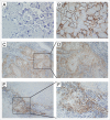
Despite its rare incidence worldwide, penile squamous cell carcinoma (PeSCC) still presents with significant morbidity and mortality due to the limited treatment options for advanced patients, especially those in developing countries. The program death-1 (PD-1)/PD-1 ligand (PD-L1) axis has been demonstrated to play an important role in tumor immune escape, and immunotherapies targeting this pathway have shown great success in certain cancer types. Here, we analyzed the expression pattern of PD-L1 in tumor cells and tumor-infiltrating lymphocytes (TILs) in PeSCC with a multi-center cohort. We found that the majority of PeSCCs (53.4%) were PD-L1-positive and that high PD-L1 expression in tumor cells was associated with a poor prognosis. Notably, PD-L1 expression in tumor cells was significantly associated with the extent of TILs and CD8+ TILs. Quantitative reverse transcriptase polymerase chain reaction (qRT-PCR) showed that PD-L1 was positively correlated with interferon-gamma (IFNγ) and CD8+ gene expression. Moreover, we defined the constitutive and inducible surface expression of PD-L1 in newly established primary PeSCC cell lines. Interestingly, two PeSCC cell lines had high intrinsic PD-L1 expression. Another cell line showed low PD-L1 expression, but the PD-L1 expression could be induced by IFNγ stimulation. Overall, our data showed that high PD-L1 expression in penile tumor cells indicated a poor prognosis. The upregulation of PD-L1 in PeSCC included both extrinsic and intrinsic mechanisms. These findings indicated that the PD-1/PD-L1 axis might be a potential therapeutic target for patients with penile squamous cell carcinoma.
Keywords: IFNγ; PD-L1; immunohistochemistry; penile cancer; penile cancer cell line; tumor-infiltrating lymphocytes.
Figures


References
-
- Hakenberg OW, Compérat EM, Minhas S, Necchi A, Protzel C, Watkin N European Association of Urology. EAU guidelines on penile cancer: 2014 update. Eur Urol 2015; 67:142-50. - PubMed
-
- Zhu Y, Gu WJ, Ye DW, Yao XD, Zhang SL, Dai B, Zhang HL, Shen YJ. External validation of nomograms for predicting cancer-specific mortality in penile cancer patients treated with definitive surgery. Chinese J Cancer 2014; 33:249-55; PMID:24559854; http://dx.doi.org/ 10.5732/cjc.013.10176 - DOI - PMC - PubMed
-
- Pandey D, Mahajan V, Kannan RR. Prognostic factors in node-positive carcinoma of the penis. J Surg Oncol 2006; 93:133-8; PMID:16425300; http://dx.doi.org/ 10.1002/jso.20414 - DOI - PubMed
-
- Rabinovich GA, Gabrilovich D, Sotomayor EM. Immunosuppressive strategies that are mediated by tumor cells. Annual Rev Immunol 2007; 25:267-96; PMID:17134371; http://dx.doi.org/ 10.1146/annurev.immunol.25.022106.141609 - DOI - PMC - PubMed
-
- Keir ME, Butte MJ, Freeman GJ, Sharpe AH. PD-1 and its ligands in tolerance and immunity. Annual Rev Immunol 2008; 26:677-704; PMID:18173375; http://dx.doi.org/ 10.1146/annurev.immunol.26.021607.090331 - DOI - PMC - PubMed
Publication types
LinkOut - more resources
Full Text Sources
Other Literature Sources
Research Materials
