. 2017 Mar 21:5:e3103.
doi: 10.7717/peerj.3103. eCollection 2017.

Evolution of the patellar sesamoid bone in mammals

Affiliations

Evolution of the patellar sesamoid bone in mammals

Mark E Samuels et al. PeerJ. .

Abstract

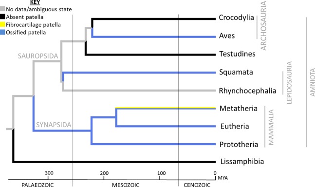
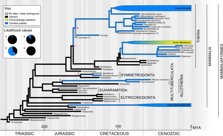
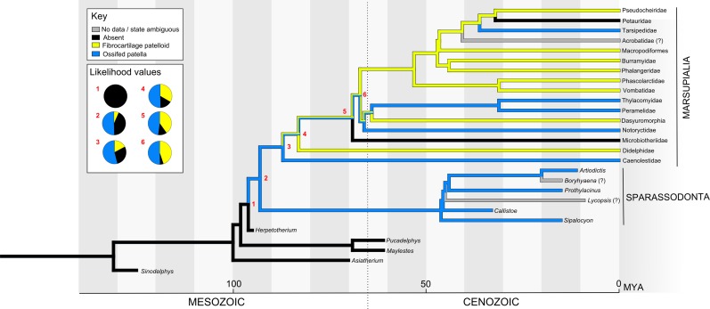
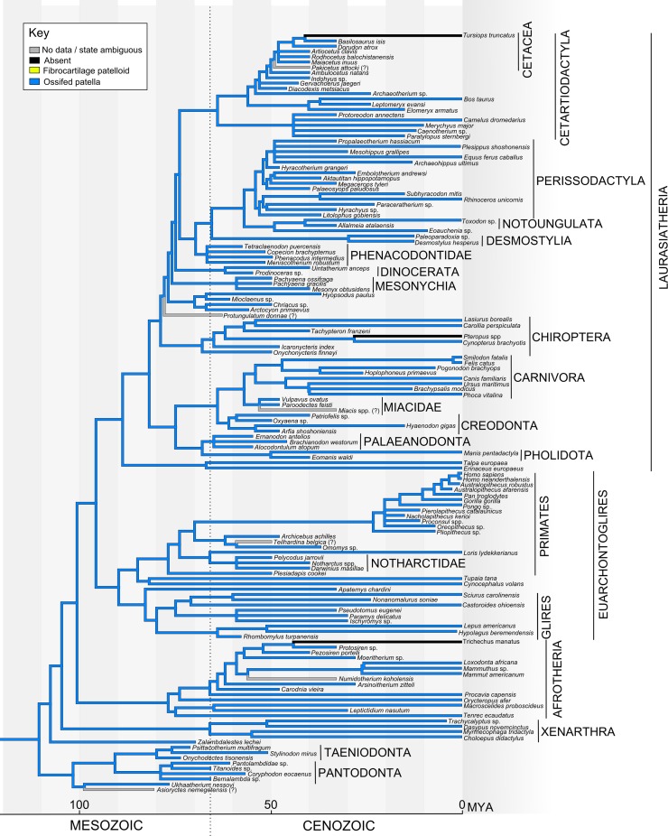
The patella is a sesamoid bone located in the major extensor tendon of the knee joint, in the hindlimb of many tetrapods. Although numerous aspects of knee morphology are ancient and conserved among most tetrapods, the evolutionary occurrence of an ossified patella is highly variable. Among extant (crown clade) groups it is found in most birds, most lizards, the monotreme mammals and almost all placental mammals, but it is absent in most marsupial mammals as well as many reptiles. Here, we integrate data from the literature and first-hand studies of fossil and recent skeletal remains to reconstruct the evolution of the mammalian patella. We infer that bony patellae most likely evolved between four and six times in crown group Mammalia: in monotremes, in the extinct multituberculates, in one or more stem-mammal genera outside of therian or eutherian mammals and up to three times in therian mammals. Furthermore, an ossified patella was lost several times in mammals, not including those with absent hindlimbs: once or more in marsupials (with some re-acquisition) and at least once in bats. Our inferences about patellar evolution in mammals are reciprocally informed by the existence of several human genetic conditions in which the patella is either absent or severely reduced. Clearly, development of the patella is under close genomic control, although its responsiveness to its mechanical environment is also important (and perhaps variable among taxa). Where a bony patella is present it plays an important role in hindlimb function, especially in resisting gravity by providing an enhanced lever system for the knee joint. Yet the evolutionary origins, persistence and modifications of a patella in diverse groups with widely varying habits and habitats-from digging to running to aquatic, small or large body sizes, bipeds or quadrupeds-remain complex and perplexing, impeding a conclusive synthesis of form, function, development and genetics across mammalian evolution. This meta-analysis takes an initial step toward such a synthesis by collating available data and elucidating areas of promising future inquiry.

Keywords: Development; Genomics; Knee; Limb; Locomotion; Osteology; Paleontology; Pathology; Phylogeny; Theria.


Conflict of interest statement

John R. Hutchinson is an Academic Editor for PeerJ.

Figures

Figure 1
Figure 1. Generalized knee showing sesamoid bones found in various mammals, although possibly no species includes all of these (patella, lunula, cyamella, fabella and parafibula).
Also shown are relevant muscles, ligaments and other anatomical elements that lie close to the sesamoids of the knee joint. The knee is in medial view and the medial collateral ligament has been removed. Illustration: Manuela Bertoni.
Figure 2
Figure 2. Generalized tetrapod with anatomical/developmental axes defined for the hindlimb: cranial/caudal (towards the head/tail, respectively), proximal/distal (toward/further from the trunk, respectively), dorsal/ventral (towards the back/belly, respectively).
Illustration: Manuela Bertoni.
Figure 3
Figure 3. Reconstruction of ancestral patellar states in Tetrapoda, showing the major extant clades.
Reconstruction was performed using Mesquite’s parsimony algorithm and unordered character states, where 0 (black) = absent patella, 1 (yellow) = soft tissue patella/patelloid and 2 (blue) = ossified patella; see “Materials and Methods” for further details. The distribution of the ossified patella among extant clades has been interpreted as three occasions of independent evolution (in Aves, Squamata and Mammalia) (Dye, 1987; Haines, 1940), a conclusion strongly reinforced by specific fossil evidence (absence or equivocality of a patella in all outgroups). Reconstruction within Mammalia is explored in more depth in Figs. 5–7. Mya, millions of years from present.
Figure 4
Figure 4. Examples of tetrapods with or without patellae.
Red arrows denote the patella. (A, B) Ornithorhynchus anatinus (Monotremata: duck-billed platypus, Redpath Museum specimen 2458). (C, D) Tachyglossus aculeatus (Monotremata: echidna, Redpath Museum specimen 2463). (E, F) D. virginiana (Metatheria: North American opossum, Redpath Museum specimen 5019). (G, H) Procavia capensis (Eutheria: Afrotheria: Cape hyrax, uncatalogued Horniman Museum Specimen, London, UK). (I) knee of patient with Meier–Gorlin Syndrome (Guernsey et al., 2010). For more images of mammalian patellae (or lack thereof in some marsupials), see Figs. S1–S3.
Figure 5
Figure 5. Ancestral state reconstruction of the patella in Mesozoic mammals (see Fig. S4 for alternative tree topology).
The main tree shows a parsimony reconstruction using unordered character states, where branch colour indicates reconstructed state. Maximum likelihood gives similar results to parsimony, and likelihood values for numbered nodes are displayed (inset). Crown Metatheria and Eutheria are further explored in Figs. 6 and 7. Our results suggest that the ossified patella has evolved at least five times within Mammaliaformes.
Figure 6
Figure 6. Ancestral state reconstruction of the patella in Metatheria and related taxa.
The main tree shows a parsimony reconstruction using unordered character states, where branch colour indicates reconstructed state. Likelihood values for the numbered nodes are shown (inset). Our results suggest that the ossified patella evolved once in Metatheria, with instances of loss and reversion (to a fibrocartilaginous patelloid and back).
Figure 7
Figure 7. Ancestral state reconstruction of the patella in Eutheria.
The main tree shows a parsimony reconstruction using unordered character states, where branch colour indicates the reconstructed state. Our results suggest that the ossified patella evolved only once within Eutheria and (as far as is currently known) has only been lost by the bat genus Pteropus (not counting groups which have lost hindlimbs, e.g. Trichechus manatus/crown Sirenia, Tursiops truncatus/crown Cetacea).

Similar articles

Cited by

References

    1. Adams RA, Thibault KM. Ontogeny and evolution of the hindlimb and calcar: assessing phylogenetic trends. In: Adams RA, Pedersen SC, editors. Ontogeny, Functional Ecology, and Evolution of Bats. Cambridge: Cambridge University Press; 2000.
    1. Aglietti P, Menchetti PPM. Biomechanics of the patellofemoral joint. In: Scuderi GR, editor. The Patella. New York: Springer; 1995.
    1. Ahlberg PE, Clack JA, Blom H. The axial skeleton of the Devonian tetrapod Ichthyostega. Nature. 2005;437(7055):137–140. doi: 10.1038/nature03893. - DOI - PubMed
    1. Alexander RM, Dimery NJ. The significance of sesamoids and retro-articular processes for the mechanics of joints. Journal of Zoology. 1985;205(3):357–371. doi: 10.1111/j.1469-7998.1985.tb05622.x. - DOI
    1. Andrews SM, Westoll TS. The postcranial skeleton of Ensthenopteron foordi whiteaves. Earth and Environmental Science Transactions of the Royal Society of Edinburgh. 1970;68(9):207–329. doi: 10.1017/s008045680001471x. - DOI

LinkOut - more resources