. 2017 Mar 21:5:e3108.
doi: 10.7717/peerj.3108. eCollection 2017.

Grape seed proanthocyanidin extract protects lymphocytes against histone-induced apoptosis

Affiliations

Grape seed proanthocyanidin extract protects lymphocytes against histone-induced apoptosis

Ping Chang et al. PeerJ. .

Abstract

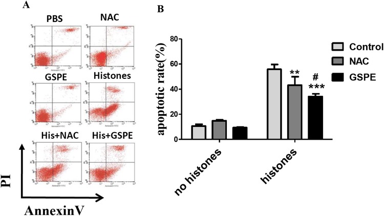
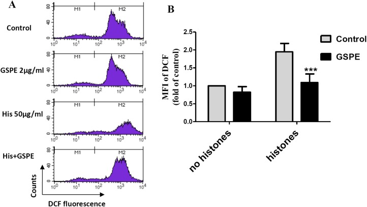
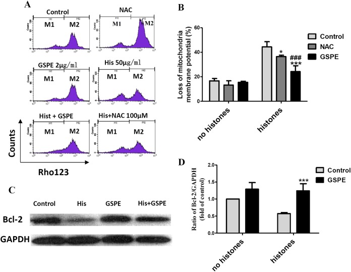
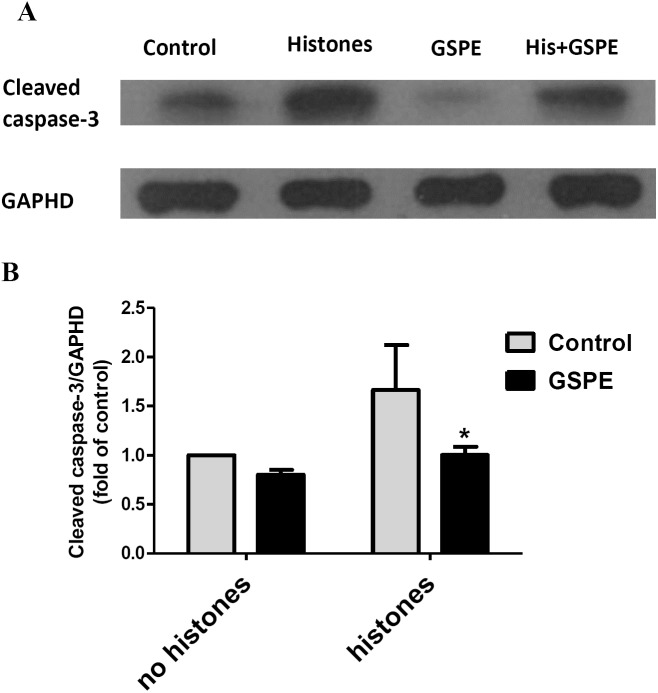
Apoptosis of lymphocytes is associated with immunosuppression and poor prognosis in sepsis. Our previous report showed that histones, nuclear proteins released from damaged or dying cells in sepsis, can mediate lymphocyte apoptosis via mitochondria damage. Grape seed proanthocyanidin extract (GSPE), a natural substance with protective properties against oxidative stress, plays a vital role in cell and mitochondria protection. We thus hypothesized that GSPE may play a protective role in histone-induced lymphocyte apoptosis through its anti-oxidative properties. In this study, we investigated the protective efficacy of GSPE on lymphocyte apoptosis induced by extracellular histones, a main contributor of death in sepsis. Human blood lymphocytes were treated with 50 μg/ml histones, 2 μg/ml GSPE, or a combination of both. A total of 100 μM N-acetylcysteine (NAC), a reactive oxygen species (ROS) inhibitor, was used as a positive control for GSPE. Apoptosis, intracellular ROS levels, mitochondrial membrane potential, Bcl-2 expression, and caspase-3 cleavage were measured. Our data clearly indicate that GSPE significantly inhibited lymphocyte apoptosis, generation of ROS, the loss of mitochondrial membrane potential, the decrease in Bcl-2 expression, and caspase-3 activation induced by extracellular histones. In conclusion, we show that GSPE has a protective effect on lymphocyte apoptosis induced by extracellular histones. This study suggests GSPE as a potential therapeutic agent that could help reduce lymphocyte apoptosis, and thus the state of immunosuppression was observed in septic patients.

Keywords: Grape seed proanthocyanidin extract; Histones; Lymphocyte apoptosis; Mitochondrial injury; Reactive oxygen species.


Conflict of interest statement

The authors declare that they have no competing interests.

Figures

Figure 1
Figure 1. Grape seed proanthocyanidin extract inhibited lymphocyte apoptosis induced by histones.
Human lymphocytes were cultured with PBS (control), GSPE (2 μg/ml), histones (His) (50 μg/ml), or histones (His) plus GSPE. NAC (100 μM) is used as a positive control of GSPE. Both GSPE and NAC were used to pre-treat cells for 2 h, then histones were added and cells cultured for an additional 2.5 h. Apoptosis were measured using Annexin V-FITC/PI double staining and flow cytometry analysis. (A) Representative pictures of lymphocytes apoptosis in indicated groups. Annexin V+ and PI− area represent early apoptosis, Annexin V+ and PI+ area represent late apoptosis. (B) Quantitative analysis of lymphocytes apoptosis. Total apoptotic lymphocytes (early and late apoptosis) were analyzed. There was a significant interaction between the effects of histones and GSPE on apoptosis, F = 15.76, Df = 2, P < 0.001. Df for error is 12. Values are presented as mean ±SD (n = 3). **P < 0.01, ***P < 0.001 vs control group treated with histones; #P < 0.05 vs NAC group treated with histones.
Figure 2
Figure 2. Grape seed proanthocyanidin extract decreased intracellular ROS formation caused by histones.
Human lymphocytes were cultured with PBS (control), histones (His) (50 μg/ml), GSPE (2 μg/ml), or histones (His) plus GSPE. Cells were pre-treated with GSPE for 2 h, then histones were added and cells cultured for an additional 2.5 h. Intracellular ROS was measured by the fluorescent probe DCFH-DA and flow cytometry analysis. (A) Representative pictures of DCF-derived fluorescence in lymphocytes in indicated groups. M1 represents the ROS− cells, M2 represents the ROS+ cells. (B) Quantitative analysis of mean fluorescence intensity (MFI) of DCF. Results were expressed as the fold of control group without histones treatment. There was a significant interaction between the effects of histones and GSPE on ROS formation, F = 10.18, Df = 1, P < 0.05. Df for error is 8. Values are presented as mean ±SD (n = 3). ***P < 0.001 vs control group treated with histones.
Figure 3
Figure 3. Grape seed proanthocyanidin extract inhibited mitochondrial damage caused by histones.
Human lymphocytes were cultured with PBS (control), NAC (100 μM), histones (His) (50 μg/ml), GSPE (2 μg/ml), histones plus GSPE, or histones (His) plus NAC. Both GSPE and NAC were used to pre-treat cells for 2 h, then histones were added and cells cultured for an additional 2.5 h. Mitochondrial membrane potential (Δψm) was detected by Rhodamine (Rho) 123 and flow cytometry analysis. (A) Representative pictures of Rho 123 fluorescence in lymphocytes in indicated groups. M1 represents the percentage of lymphocytes with Δψm loss, M2 represents the percentage of lymphocytes without Δψm loss. (B) Bar graph showing differences in the percentage of cells with Δψm loss. There was a significant interaction between the effects of histones and GSPE on Δψm loss, F = 15.31, Df = 1, P < 0.001. Df for error is 12. Values were presented as mean ±SD (n = 3). *P < 0.05, ***P < 0.001 vs control group treated with histones; ###P < 0.001 vs NAC group treated with histones. (C) Representative blots of Bcl-2 expression in lymphocytes in indicated groups. Human lymphocytes were cultured with PBS (control), histone (His) (50 μg/ml), GSPE (2 μg/ml), or histones (His) plus GSPE. GSPE was used to pre-treat cells for 2 h, then histones were added and cultured for 2.5 h. Bcl-2 expression was evaluated by western blotting. GAPDH was used as a loading control to normalize data for differences in the amount of total proteins loaded per lane. (D) Quantification of Bcl-2/GAPDH expression ratio. Densitometric analysis of relative bcl-2 band intensities were used to compare groups. Results were expressed as fold change compared to control group without histones treatment. The main effects of histones and GSPE are significant. For histones, F = 8.22, Df = 1, P < 0.05, for GSPE, F = 33.12, Df = 1, P < 0.001. Df for error is 8. Values are presented as mean ±SD (n = 3). ***P < 0.001 vs control group treated with histones.
Figure 4
Figure 4. Grape seed proanthocyanidin extract inhibited caspase-3 activation induced by histones.
Human lymphocytes were cultured with PBS (control), histones (His) (50 μg/ml), GSPE (2 μg/ml), or histones (His) plus GSPE. GSPE was used to pre-treat cells for 2 h, then histones were added and cells cultured for an additional 2.5 h. Control cells were treated with PBS. Cleaved caspase-3 were detected by western blotting. GAPDH was used as a loading control to normalize data for differences in the amount of total proteins loaded per lane. (A) Representative blots of cleaved caspase-3 expression for each group are shown. Cleaved caspase-3 represents the activated form of caspase-3. (B) Quantification of cleaved caspase-3/GAPDH expression ratio. Densitometric analysis of relative cleaved caspase-3 band intensities were used to compare groups. Results were expressed as the fold change compared to control group without histones treatment. The main effects of histones and GSPE are significant. For histones, F = 10.48, Df = 1, P < 0.05, for GSPE, F = 10.32, Df = 1, P < 0.05. Df for error is 8. Values are presented as mean ±SD (n = 3). *P < 0.05 vs control group treated with histones.

Similar articles

Cited by

References

    1. Ahmad M. Protective effects of curcumin against lithium–pilocarpine induced status epilepticus, cognitive dysfunction and oxidative stress in young rats. Saudi Journal of Biological Sciences. 2013;20(2):155–162. doi: 10.1016/j.sjbs.2013.01.002. - DOI - PMC - PubMed
    1. Ahn SH, Kim HJ, Jeong I, Hong YJ, Kim MJ, Rhie DJ, Jo YH, Hahn SJ, Yoon SH. Grape seed proanthocyanidin extract inhibits glutamate-induced cell death through inhibition of calcium signals and nitric oxide formation in cultured rat hippocampal neurons. BMC Neuroscience. 2011;12(1):78. doi: 10.1186/1471-2202-12-78. - DOI - PMC - PubMed
    1. Bagchi D, Sen CK, Ray SD, Das DK, Bagchi M, Preuss HG, Vinson JA. Molecular mechanisms of cardioprotection by a novel grape seed proanthocyanidin extract. Mutation Research. 2003;523–524:87–97. doi: 10.1016/s0027-5107(02)00324-x. - DOI - PubMed
    1. Cepinskas G, Wilson JX. Inflammatory response in microvascular endothelium in sepsis: role of oxidants. Journal of Clinical Biochemistry and Nutrition. 2008;42(3):175–184. doi: 10.3164/jcbn.2008026. - DOI - PMC - PubMed
    1. Demirkaya E, Avci A, Kesik V, Karslioglu Y, Oztas E, Kismet E, Gokcay E, Durak I, Koseoglu V. Cardioprotective roles of aged garlic extract, grape seed proanthocyanidin, and hazelnut on doxorubicin-induced cardiotoxicity. Canadian Journal of Physiology and Pharmacology. 2009;87(8):633–640. doi: 10.1139/y09-051. - DOI - PubMed

LinkOut - more resources