. 2017 Feb 13:4:204-212.
doi: 10.1016/j.omtm.2017.01.004. eCollection 2017 Mar 17.

Chronic Enzyme Replacement to the Brain of a Late Infantile Neuronal Ceroid Lipofuscinosis Mouse Has Differential Effects on Phenotypes of Disease

Affiliations

Chronic Enzyme Replacement to the Brain of a Late Infantile Neuronal Ceroid Lipofuscinosis Mouse Has Differential Effects on Phenotypes of Disease

Jennifer A Wiseman et al. Mol Ther Methods Clin Dev. .

Abstract

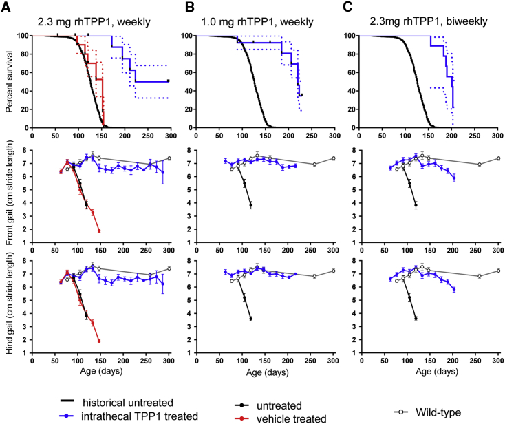
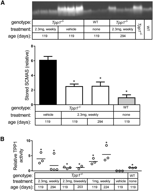
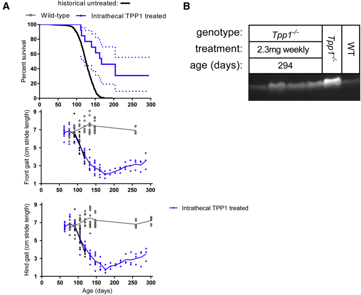
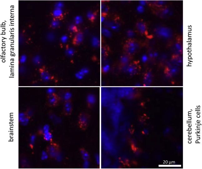
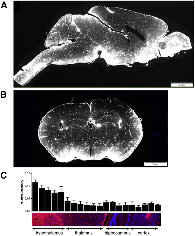
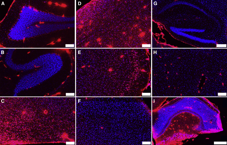
Late infantile neuronal ceroid lipofuscinosis (LINCL) is a fatal inherited neurodegenerative disease caused by loss of lysosomal protease tripeptidyl peptidase 1 (TPP1). We have investigated the effects of chronic intrathecal (IT) administration using enzyme replacement therapy (ERT) to the brain of an LINCL mouse model, in which locomotor function declines dramatically prior to early death. Median lifespan was significantly extended from 126 days to >259 days when chronic IT treatment was initiated before the onset of disease. While treated animals lived longer and showed little sign of locomotor dysfunction as measured by stride length, some or all (depending on regimen) still died prematurely. One explanation is that cerebrospinal fluid (CSF)-mediated delivery may not deliver TPP1 to all brain regions. Morphological studies support this, showing delivery of TPP1 to ventral, but not deeper and dorsal regions. When IT treatment is initiated in severely affected LINCL mice, lifespan was extended modestly in most but dramatically extended in approximately one-third of the cohort. Treatment improved locomotor function in these severely compromised animals after it had declined to the point at which animals normally die. This indicates that some pathology in LINCL is reversible and does not simply reflect neuronal death.

Keywords: chronic; enzyme replacement therapy; intrathecal; neuronal ceroid lipofuscinosis; tripeptidyl peptidase 1.


Figures

Figure 1
Figure 1
Chronic IT Enzyme Replacement Therapy of Asymptomatic Animals Treatment was initiated at 42 days of age. Dashed lines in survival curves are SE; gait error bars are SEM. Treatment groups were compared with untreated animals using log-rank (Mantel-Cox) tests. (A) Survival of animals treated weekly with 2.3 mg rhTPP1 (n = 13 animals) was significantly increased (p < 0.0001) from vehicle (aCSF) alone (n = 10) or untreated animals. (B) Survival of animals treated with a weekly administration of 1 mg rhTPP1 (n = 12) was significantly (p < 0.0001) increased. (C) Survival of animals treated with a biweekly administration of 2.3 mg rhTPP1 (n = 12) was significantly (p < 0.0001) increased.
Figure 2
Figure 2
Storage Material and TPP1 Activity Significance of changes in SCMAS and TPP1 activity relative to vehicle-treated LINCL mice was calculated by fitting data to a linear model, as described in the Materials and Methods. (A) Detection of SCMAS in isolated storage material by immunoblotting in animals treated with a weekly administration of 2.3 mg rhTPP1 and controls. Samples from 16-week-old wild-type and LINCL animals are shown as reference standards. Analysis of older wild-type animals indicated that there were no detectable age-related changes in SCMAS levels (data not shown). Relative SCMAS levels obtained from multiple loadings (4 and 8 μg protein equivalents of original homogenate) were normalized to average signal obtained from 17-week-old wild-type animals; error bars show SEM (n = 3 per treatment group). Significance of change compared to vehicle-treated LINCL mice, *p < 0.05/3 (i.e., 0.05 after Bonferroni correction for three individual comparisons). (B) TPP1 activities measured in brain, 24 hours after administration of the final dose. Data are expressed relative to average TPP1 activity in 17-week-old wild-type animals. Significance of change compared to vehicle treated animals, *p < 0.05/7 (i.e., 0.05 after Bonferroni correction for seven individual comparisons).
Figure 3
Figure 3
IT ERT in Late-Stage Disease Weekly administration of 2.3 mg rhTPP1 was initiated at 105 days of age (n = 12 animals). (A) Survival and gait analysis data. Dashed lines in survival plot indicate SE. Survival of treated animals was significantly increased compared to untreated animals (p < 0.0001, log-rank [Mantel-Cox] test). Stride length is plotted for each individual animal to demonstrate variability. Linear regression of stride length data from 175 to 287 days indicates that the increase in stride length observed after 175 days is highly significant (hind gait, slope = 0.015 ± 0.002, p < 0.0001; front gait, slope = 0.013 ± 0.002, p < 0.0001). (B) SCMAS in LINCL mice at 42 weeks of age. The ∼16-week-old wild-type and LINCL animals are shown as reference standards.
Figure 4
Figure 4
Intracellular Distribution of Fluorescent TPP1 Wild-type mice were treated with 1 mg Alexa-647-labeled rhTPP1 via IT administration. Then 18 hr later, animals were killed, and brains were analyzed by confocal microscopy to measure the distribution of fluorescently labeled TPP1. Images were captured using a 40× objective. Blue, DAPI; red, Alexa-647 rhTPP1. Note that images were chosen for each brain region to illustrate intracellular distribution and are not meant to be representative of each respective brain region as a whole.
Figure 5
Figure 5
Distribution of Fluorescent TPP1 (A–C) Wild-type mice (n = 3) were treated with Alexa-647-labeled rhTPP1 via IT administration. Then 18 hr later, animals were killed, and brains were analyzed by confocal microscopy to measure the distribution of fluorescently labeled rhTPP1. (A) Sagittal section, (B) coronal section, and (C) quantitation of relative fluorescent rhTPP1 signal through a dorsal-ventral section are shown (n = 3 animals, error bars depict SEM). Images were captured using a 20× objective. Blue, DAPI; red, Alexa-647 rhTPP1.
Figure 6
Figure 6
Distribution of Fluorescent rhTPP1 in Defined Brain Regions in Sagittal Section See Figure 5 for details. Color ranges in all panels are identical. (A) Cerebellum, lobe VIII; (B) cerebellum, lobe I; (C) hypothalamus; (D) brainstem; (E) anterior cerebral cortex; (F) posterior cerebral cortex; (G) hippocampus; (H) thalamus; and (I) olfactory bulb are shown. Scale bars represent 100 μm except in (G) (200 μm) and (I) (500 μm). Blue, DAPI; red, Alexa-647-labeled rhTPP1.

References

    1. Sleat D.E., Donnelly R.J., Lackland H., Liu C.G., Sohar I., Pullarkat R.K., Lobel P. Association of mutations in a lysosomal protein with classical late-infantile neuronal ceroid lipofuscinosis. Science. 1997;277:1802–1805. - PubMed
    1. Mole S.E., Williams R.E., Goebel H.H. Oxford University Press; Oxford: 2011. The Neuronal Ceroid Lipofuscinoses (Batten Disease)
    1. Hollak C.E., Wijburg F.A. Treatment of lysosomal storage disorders: successes and challenges. J. Inherit. Metab. Dis. 2014;37:587–598. - PubMed
    1. Nickel M., Jacoby D., Lezius S., Down M., Simonati A., Genter F., Wittes J., Kohlschütter A., Schulz A. Natural history of CLN2 disease: quantitative assessment of disease characteristics and rate of progression. Neuropediatrics. 2016;47:FV04-03.
    1. Schulz A., Specchio N., Gissen P., de los Reyes E., Williams R., Cahan H., Genter F., Jacoby D. Intracerebroventricular Cerliponase Alfa (BMN 190) in children with CLN2 disease: interim results from a phase 1/2, open-label, dose-escalation study. Neuropediatrics. 2016;47:FV02-06.

LinkOut - more resources