. 2016 Dec 14:4:55-66.
doi: 10.1016/j.omto.2016.12.001. eCollection 2017 Mar 17.

OvAd1, a Novel, Potent, and Selective Chimeric Oncolytic Virus Developed for Ovarian Cancer by 3D-Directed Evolution

Affiliations

OvAd1, a Novel, Potent, and Selective Chimeric Oncolytic Virus Developed for Ovarian Cancer by 3D-Directed Evolution

Irene Kuhn et al. Mol Ther Oncolytics. .

Abstract

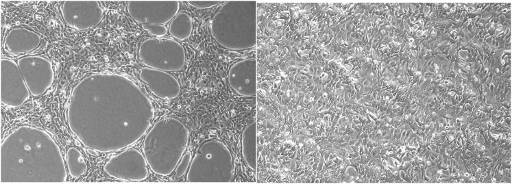
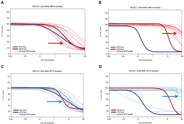
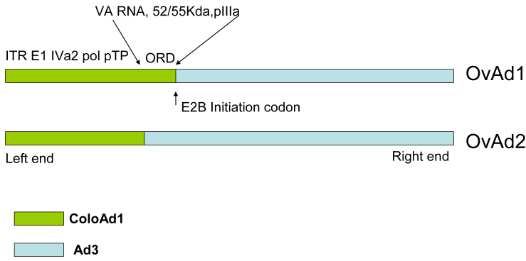
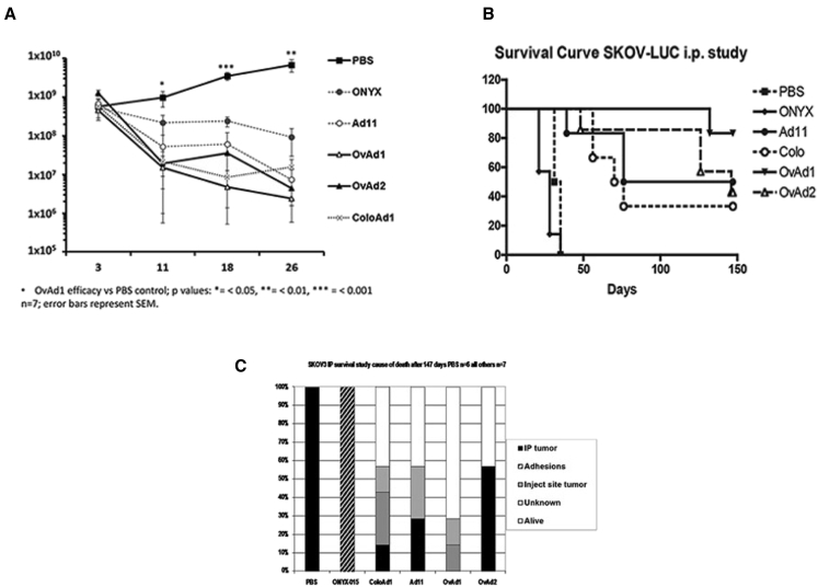
Effective therapeutics for ovarian cancer continue to be urgently needed, particularly for chemotherapy-resistant cases. Here we present both a 3D-Matrigel culture-based expansion of our directed evolution method for generation of oncolytic virotherapies and two promising ovarian-cancer targeted oncolytic viruses, OvAd1 and OvAd2. OvAd1 was developed using Matrigel cell cultures, whereas OvAd2 was developed in parallel using traditional monolayer tissue culture methods. Both viruses are potent against a panel of platinum-resistant ovarian cancer cell lines and are attenuated on normal cells in vitro, resulting in therapeutic windows of ∼200-fold. We observed two benefits of the use of Matrigel-based cultures for directed evolution of these oncolytics: (1) use of Matrigel generated a bioselected pool that was more strongly attenuated on normal cells while retaining its potency against ovarian cancer cells, and (2) in an ovarian carcinomatosis model, the Matrigel-derived virus OvAd1 suppressed all tumor growth while the non-Matrigel-derived virus was 50% effective. Neither virus stimulated formation of peritoneal adhesions as seen for Ad5-based therapies. Consequently, these viruses are novel candidates for development as new effective treatments for aggressive ovarian cancer.

Keywords: 3D cell culture; Matrigel; adenovirus; directed evolution; oncolytic; ovarian cancer; replicating; virotherapy.


Figures

Figure 1
Figure 1
SKOV3 Cells Grown on Matrigel Acquire Substratum-Dependent Morphology SKOV3 cells grown on GFR-Matrigel (left) compared with those grown by traditional method on tissue culture plastic (right) display distinct morphologies.
Figure 2
Figure 2
AIEX Analysis of SP10 and SM10 Identifies Ad Serotypes in Each Pool and Allows Accurate Viral Particle Counts The bioselected pools, SM10 and SP10, were analyzed by AIEX chromatography. The elution positions of adenoviral peaks are characteristic for each adenoviral serotype (Figure S2) and are evident in the chromatograms as the narrow peaks superimposed on the broader protein peaks (discussed in Kuhn et al.19) contributed by the SKOV3 culture medium. The position of each narrow peak in the chromatograms of the passaged viral pools indicate that SP10 (dark green chromatogram) is predominantly composed of Ad3-related viruses, whereas SM10 (blue chromatogram) contains both Ad3 and Ad11p-related viruses.
Figure 3
Figure 3
Assessing Tumor Specificity: Potency of the SM10 and SP10 Pools and Viral Isolates on SKOV3 versus HUVEC Monolayers (A) Potency of SM10 pool isolates assessed on monolayers of SKOV3 cells. The SP10 and SM10 pools were included in all MTS analyses as references to allow comparison between assays. Results at 6 dpi are presented. The SM10 and SP10 pools as well as the 12 isolates of the SM10 pool had similar potencies (within a 1-log range) against SKOV3 cells. The red arrow indicates the clonal isolate of SM10 that was renamed OvAd1. (B) The same viral isolates and pools as in (A) were assessed in parallel on HUVEC monolayers; endpoints read at 10 dpi (the HUVEC assay required more days than the SKOV3 assay to reveal any killing of HUVECs by the 12 SM10 isolates). The SM10 pool was about 2 logs more attenuated on HUVECs than was the SP10 pool, and the SM10 pool isolates were 2–3 logs more attenuated on HUVECs than was the SP10 pool. The red arrow indicates the clonal isolate of SM10 that was renamed OvAd1. (C) Potency of SP10 pool isolates assessed on monolayers of SKOV3 cells, along with the SM10 and the SP10 pools; results at 6 dpi are presented. As in (A), SM10 and SP10 pools had very similar potencies on SKOV3, and the SP10 isolates were within about a 1-log range of one another and of the pools. The blue arrow indicates the clonal isolate of SP10 that was renamed OvAd2. (D) The same viral isolates and pools as in (C) were analyzed in parallel on HUVECs. Results at 7 dpi are presented. The SP10 pool and two SP10 isolates are 3 or more logs more potent against HUVEC than the SM10 pool, indicating that the SP10 pool and these two isolates are at best weakly tumor selective. The remaining 10 isolates of the SP10 pool appear to be tumor selective. The blue arrow indicates the clonal isolate of SP10 that was renamed OvAd2.
Figure 4
Figure 4
OvAd1 and OvAd2 Are Chimeras of ColoAd1 and Ad3
Figure 5
Figure 5
OvAd1 and OvAd2 Proved Effective in Mouse Model of Ovarian Cancer and Did Not Generate Peritoneal Adhesions Ovarian carcinomatosis tumors were established by administration of 5 × 106 SKOV-3.Luc cells on day 0 (t0) into the peritoneal cavity and randomly assigned to seven groups of seven mice each. At days 3, 5, and 7, mice were administered PBS, ONYX-015, Ad11p, OvAd1, or OvAd2 via a 0.5-mL i.p. injection. All virus-treated mice received a dose of 5 × 1010 viral particles (vp) per injection for a total of 1.5 × 1011 vp/mouse. (A) During the first 26 days, tumor burden was followed by luciferase imaging, and (B) survival was followed out to 147 days. (C) Autopsy observations of pathological changes contributing to mouse mortality.

Similar articles

Cited by

References

    1. Lowe K.A., Chia V.M., Taylor A., O’Malley C., Kelsh M., Mohamed M., Mowat F.S., Goff B. An international assessment of ovarian cancer incidence and mortality. Gynecol. Oncol. 2013;130:107–114. - PubMed
    1. English D.P., Menderes G., Black J., Schwab C.L., Santin A.D. Molecular diagnosis and molecular profiling to detect treatment-resistant ovarian cancer. Expert Rev. Mol. Diagn. 2016;16:769–782. - PubMed
    1. Buller R.E., Runnebaum I.B., Karlan B.Y., Horowitz J.A., Shahin M., Buekers T., Petrauskas S., Kreienberg R., Slamon D., Pegram M. A phase I/II trial of rAd/p53 (SCH 58500) gene replacement in recurrent ovarian cancer. Cancer Gene Ther. 2002;9:553–566. - PubMed
    1. Aghi M., Martuza R.L. Oncolytic viral therapies—the clinical experience. Oncogene. 2005;24:7802–7816. - PubMed
    1. Vasey P.A., Shulman L.N., Campos S., Davis J., Gore M., Johnston S., Kirn D.H., O’Neill V., Siddiqui N., Seiden M.V., Kaye S.B. Phase I trial of intraperitoneal injection of the E1B-55-kd-gene-deleted adenovirus ONYX-015 (dl1520) given on days 1 through 5 every 3 weeks in patients with recurrent/refractory epithelial ovarian cancer. J. Clin. Oncol. 2002;20:1562–1569. - PubMed