. 2016 Dec 31:4:77-86.
doi: 10.1016/j.omto.2016.12.004. eCollection 2017 Mar 17.

Oncolytic Adenoviruses Armed with Tumor Necrosis Factor Alpha and Interleukin-2 Enable Successful Adoptive Cell Therapy

Affiliations

Oncolytic Adenoviruses Armed with Tumor Necrosis Factor Alpha and Interleukin-2 Enable Successful Adoptive Cell Therapy

Riikka Havunen et al. Mol Ther Oncolytics. .

Abstract

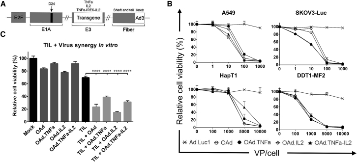
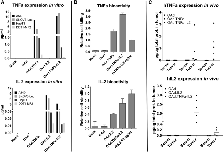
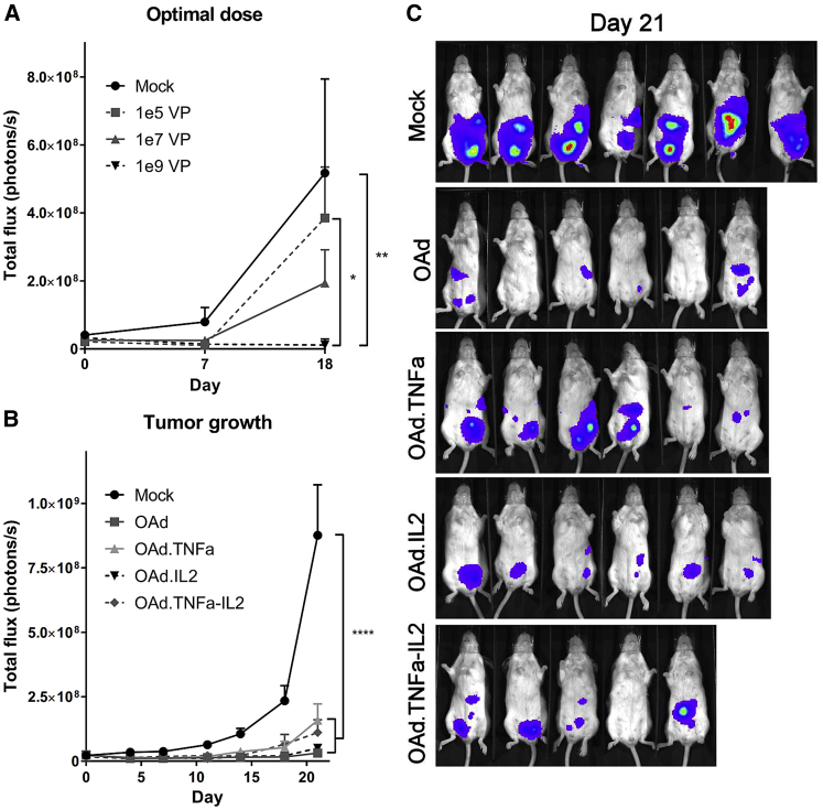
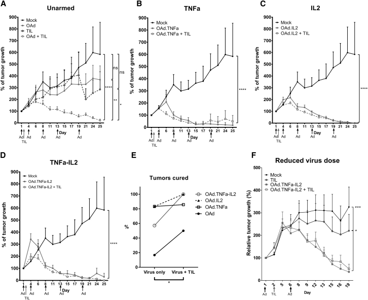
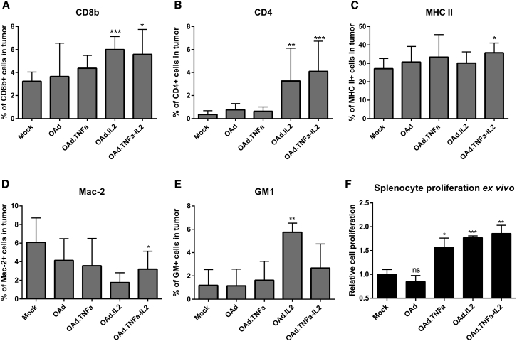
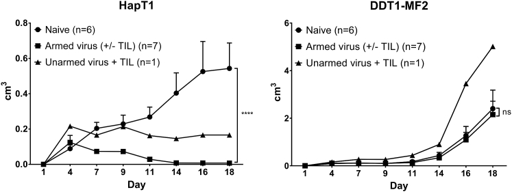
Adoptive cell therapy holds much promise in the treatment of cancer but results in solid tumors have been modest. The notable exception is tumor-infiltrating lymphocyte (TIL) therapy of melanoma, but this approach only works with high-dose preconditioning chemotherapy and systemic interleukin (IL)-2 postconditioning, both of which are associated with toxicities. To improve and broaden the applicability of adoptive cell transfer, we constructed oncolytic adenoviruses coding for human IL-2 (hIL2), tumor necrosis factor alpha (TNF-α), or both. The viruses showed potent antitumor efficacy against human tumors in immunocompromised severe combined immunodeficiency (SCID) mice. In immunocompetent Syrian hamsters, we combined the viruses with TIL transfer and were able to cure 100% of the animals. Cured animals were protected against tumor re-challenge, indicating a memory response. Arming with IL-2 and TNF-α increased the frequency of both CD4+ and CD8+ TILs in vivo and augmented splenocyte proliferation ex vivo, suggesting that the cytokines were important for T cell persistence and proliferation. Cytokine expression was limited to tumors and treatment-related signs of systemic toxicity were absent, suggesting safety. To conclude, cytokine-armed oncolytic adenoviruses enhanced adoptive cell therapy by favorable alteration of the tumor microenvironment. A clinical trial is in progress to study the utility of Ad5/3-E2F-d24-hTNFa-IRES-hIL2 (TILT-123) in human patients with cancer.

Keywords: TNF; adenovirus; cytokines; interleukin 2; tumor-infiltrating lymphocytes.


Figures

Figure 1
Figure 1
Oncolytic Activity of Adenoviral Vectors in Human and Hamster Cancer Cell Lines (A) A schematic presentation of chimeric oncolytic adenovirus with E2F promoter; 24-base-pair deletion in E1A; human TNF-α, IL-2, or TNF-α-IRES-IL2 inserted in the E3 region; and an Ad3 serotype knob in the Ad5 fiber. (B) Oncolytic activity of the viruses was shown in human lung adenocarcinoma (A549) and luciferase-expressing ovarian carcinoma (SKOV3-Luc) as well as in hamster pancreatic cancer (HapT1) and leiomyosarcoma (DDT1-MF2). The cells were incubated with the viruses for 3 days (A549 and DDT1-MF2), 5 days (SKOV3-Luc), or 6 days (HapT1) before determining cell viability. (C) Cell-killing efficacy was enhanced when combining viruses with HapT1-specific TILs. The cells were incubated 72 hr with 5,000 VPs and 24 hr with TILs. Means ± SEM are shown (n = 8). Statistical differences were evaluated with one-way ANOVA. ****p ≤ 0.0001. Ad.Luc1, replication-deficient Ad5/3-Luc1; OAd, Ad5/3-E2F-d24; OAd.TNFa, Ad5/3-E2F-d24-hTNFa; OAd.IL2, Ad5/3-E2F-d24-hIL2; OAd.TNFa-IL2, Ad5/3-E2F-d24-hTNFa-IRES-hIL2.
Figure 2
Figure 2
Biologically Active Cytokines Are Produced from Human and Hamster Cell Lines and Expressed Locally In Vivo (A) Human and hamster cell lines were incubated for 72 hr with 1,000 or 5,000 VPs per cell, respectively. Cytokine concentrations were measured from cell culture supernatants with a cytometric bead array. (B) Indicator cell line L929 was used in the cell-killing assay to confirm the biological activity of TNF-α, while mouse T cell line CTLL-2 was utilized in the IL-2-induced cell proliferation assay. The samples were derived from virus-infected HapT1 supernatants. Means ± SD are shown. (C) HapT1 tumors were injected with 1 × 108 VPs and collected together with blood after 48 hr. Cytokine concentrations were measured with a cytometric bead array and normalized against total protein concentration of the sample. Horizontal lines indicate mean values. OAd, Ad5/3-E2F-d24; OAd.TNFa, Ad5/3-E2F-d24-hTNFa; OAd.IL2, Ad5/3-E2F-d24-hIL2; OAd.TNFa-IL2, Ad5/3-E2F-d24-hTNFa-IRES-hIL2.
Figure 3
Figure 3
Armed Oncolytic Adenoviruses Show Dose-Dependent Antitumor Efficacy in Immunocompromised Mice (A–C) SCID mice bearing orthotopic luciferase-expressing ovarian carcinoma SKOV3-Luc were treated with (A) different doses of Ad5/3-E2F-d24-hTNFa-IRES-hIL2 (n = 3) and with (B and C) all the viruses with selected dose (1 × 109 VPs/animal, n = 5–7). Means ± SEM are shown. Statistical differences were evaluated with two-way ANOVA. *p ≤ 0.05; **p ≤ 0.01; ****p ≤ 0.0001. (C) Bioluminescent images from day 21 were merged with photographs. The color scale from violet to red represents luminescence intensity from 1 × 106 to 1 × 108 p/s/cm2/sr, respectively. OAd, Ad5/3-E2F-d24; OAd.TNFa, Ad5/3-E2F-d24-hTNFa; OAd.IL2, Ad5/3-E2F-d24-hIL2; OAd.TNFa-IL2, Ad5/3-E2F-d24-hTNFa-IRES-hIL2.
Figure 4
Figure 4
Armed Oncolytic Adenoviruses Improve the Curative Efficacy of Adoptive TIL Therapy in Immunocompetent Syrian Hamsters (A–D) Syrian hamsters with established subcutaneous HapT1 tumors were treated five times with 1 × 109 VPs and once with 4 × 105 freshly expanded TILs, both injected intratumorally (n = 6–7). Tumors lacking neoplastic cells were considered cured. Statistical differences were evaluated with a linear mixed-effects model. (E) Complete tumor remission was evaluated before animals were euthanized or in unclear cases after histopathological analysis. The effect of TILs on cure rates was evaluated with the Wilcoxon signed-rank test. (F) The experiment was repeated with a reduced virus dose (1 × 108 VPs on days 1 and 8, n = 5–6). Differences in antitumor efficacy were analyzed with two-way ANOVA. *p ≤ 0.05; **p ≤ 0.01; ***p ≤ 0.001; ****p ≤ 0.0001. ns, non-significant; OAd, Ad5/3-E2F-d24; OAd.TNFa, Ad5/3-E2F-d24-hTNFa; OAd.IL2, Ad5/3-E2F-d24-hIL2; OAd.TNFa-IL2, Ad5/3-E2F-d24-hTNFa-IRES-hIL2.
Figure 5
Figure 5
Adenovirus Treatments Induce Beneficial Immunological Changes in Tumor Microenvironment (A–E) The following cells were stained from endpoint tumors with fluorochrome-conjugated antibodies and analyzed by flow cytometry: (A) CD8+, (B) CD4+, (C) MHC II+, (D) Mac-2+, and (E) GM1+. Because of the great number of cured tumors, groups with or without TILs were combined. Means ± SD are shown (n = 3–9). Differences in cell percentages were analyzed with the Student’s t test. (F) Splenocyte proliferation was determined by counting the cells. Because TILs did not affect the proliferation, groups with or without TILs were combined for simplification. Means ± SEM are shown (n = 4). Statistical significance was calculated with the non-parametric Mann-Whitney test. *p ≤ 0.05; **p ≤ 0.01; ***p ≤ 0.001. ns, non-significant; OAd, Ad5/3-E2F-d24; OAd.TNFa, Ad5/3-E2F-d24-hTNFa; OAd.IL2, Ad5/3-E2F-d24-hIL2; OAd.TNFa-IL2, Ad5/3-E2F-d24-hTNFa-IRES-hIL2.
Figure 6
Figure 6
Cytokine-Armed Virus Treatments Induce Tumor-Specific Immunological Memory Hamsters previously cured of HapT1 tumors were re-challenged with the same HapT1 tumor cells or with a distinct cell line (DDT1-MF2). Hamsters previously treated with cytokine-armed viruses completely rejected the re-introduced HapT1 tumors, whereas the hamster treated with unarmed virus had a stable condition. Means ± SEM are presented. ****p ≤ 0.0001. ns, non-significant.

References

    1. Rosenberg S.A., Yang J.C., Sherry R.M., Kammula U.S., Hughes M.S., Phan G.Q., Citrin D.E., Restifo N.P., Robbins P.F., Wunderlich J.R. Durable complete responses in heavily pretreated patients with metastatic melanoma using T-cell transfer immunotherapy. Clin. Cancer Res. 2011;17:4550–4557. - PMC - PubMed
    1. Larkin J., Chiarion-Sileni V., Gonzalez R., Grob J.J., Cowey C.L., Lao C.D., Schadendorf D., Dummer R., Smylie M., Rutkowski P. Combined nivolumab and ipilimumab or monotherapy in untreated melanoma. N. Engl. J. Med. 2015;373:23–34. - PMC - PubMed
    1. Andtbacka R.H., Kaufman H.L., Collichio F., Amatruda T., Senzer N., Chesney J., Delman K.A., Spitler L.E., Puzanov I., Agarwala S.S. Talimogene laherparepvec improves durable response rate in patients with advanced melanoma. J. Clin. Oncol. 2015;33:2780–2788. - PubMed
    1. Li H., Wang C., Yu J., Cao S., Wei F., Zhang W., Han Y., Ren X.B. Dendritic cell-activated cytokine-induced killer cells enhance the anti-tumor effect of chemotherapy on non-small cell lung cancer in patients after surgery. Cytotherapy. 2009;11:1076–1083. - PubMed
    1. Zhao X., Zhang Z., Li H., Huang J., Yang S., Xie T., Huang L., Yue D., Xu L., Wang L. Cytokine induced killer cell-based immunotherapies in patients with different stages of renal cell carcinoma. Cancer Lett. 2015;362:192–198. - PubMed