. 2017 Jun;26(6):1171-1181.
doi: 10.1002/pro.3167. Epub 2017 Apr 2.

NMR solution structure of the RED subdomain of the Sleeping Beauty transposase

Affiliations

NMR solution structure of the RED subdomain of the Sleeping Beauty transposase

Tatiana A Konnova et al. Protein Sci. 2017 Jun.

Abstract

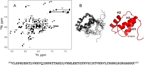
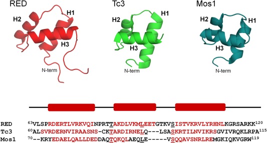
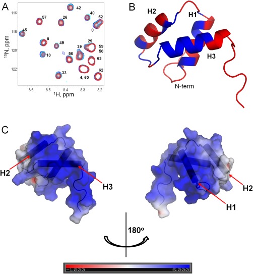
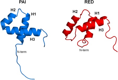
DNA transposons can be employed for stable gene transfer in vertebrates. The Sleeping Beauty (SB) DNA transposon has been recently adapted for human application and is being evaluated in clinical trials, however its molecular mechanism is not clear. SB transposition is catalyzed by the transposase enzyme, which is a multi-domain protein containing the catalytic and the DNA-binding domains. The DNA-binding domain of the SB transposase contains two structurally independent subdomains, PAI and RED. Recently, the structures of the catalytic domain and the PAI subdomain have been determined, however no structural information on the RED subdomain and its interactions with DNA has been available. Here, we used NMR spectroscopy to determine the solution structure of the RED subdomain and characterize its interactions with the transposon DNA.

Keywords: DNA binding; NMR spectroscopy; RED subdomain; SB transposase; folding; structure.


Figures

Figure 1
Figure 1
Folding of the RED subdomain in the presence of crowding. Far UV CD and [1H,15N]‐HSQC NMR spectra of the RED subdomain in the presence of PEG 6000 (A,B) and Ficoll‐70 (C,D) were collected in 20 mM aqueous MES buffer at pH 5.0. Increasing the concentration of crowders does not induce a significant amount of alpha‐helical structure. Arrows indicate increasing concentrations of crowders.
Figure 2
Figure 2
Folding of the RED subdomain in the presence of different salts. Far UV CD spectra of the RED subdomain collected in 20 mM aqueous MES buffer at pH 5.0 in the presence of up to 800 mM of NaClO4 (A), NaCl (B), KCl (C), and Na2SO4 (D). Arrows indicate increasing concentrations of salts. The largest content of alpha‐helical is observed in the presence of Na2SO4.
Figure 3
Figure 3
NMR solution structure of the RED subdomain. (A) Assigned [15N‐1H]‐HSQC spectrum of the RED subdomain. The spectrum was recorded in 20 mM aqueous (5% D2O/95% H2O) MES buffer at pH 5.0 in the presence of 650 mM Na2SO4. The amino acid sequence of the RED subdomain, containing residues 63–120 of the original SB transposase sequence,7 is shown below the spectrum for reference. (B) NMR solution structure of the RED subdomain. Cα traces of superimposed 10 lowest energy structures (left) and the cartoon representation of the representative structure of RED (right) are shown. The identified alpha helices are labeled as H1, H2, and H3.
Figure 4
Figure 4
Structural alignment of the RED subdomain of the SB transposase and C‐terminal DNA‐binding subdomains of Tc3 (pdb code 1U7826) and Mos1 (pdb code 3HOS15) transposases. Protein structures were superimposed and then merely shifted along x (horizontal) axis. Structural alignment was done by using PROMALS3D multiple sequence and structure alignment server.27 The RMSD over backbone Cα atoms between the RED subdomain and the corresponding Tc3 or Mos1 subdomain structures is 3.5 Å and 3.7 Å, respectively. The three alpha helices are labeled as H1, H2, and H3. The helix‐turn helix motif in Tc3 and Mos1 subdomains is formed by helices H2 and H3. The amino acid sequence alignment according to the correspondence of the three structures shows low amino acid sequence similarity between the RED subdomain and the corresponding subdomains of Tc3 and Mos1 transposases, and the only three residues in conserved positions are underlined. Residues that form alpha helices are shown in red. The position of helices in the RED subdomain is also indicated by red rectangles above the amino acid sequence.
Figure 5
Figure 5
Binding of the RED subdomain to DNA. (A) Section of the [15N,1H]‐HSQC spectrum of the pure RED subdomain (blue) and of the RED subdomain in the presence of 1:0.5 (cyan) and 1:1 (red) molar ratio of DNA sequence corresponding to the outer transposase binding site on the left ITR (Lo). The spectra were collected in aqueous (5% D2O/95% H2O) 20 mM MES buffer at pH 5.0 in the presence of 650 mM Na2SO4. (B) Cartoon structure of the RED subdomain shows residues affected by the binding to the Lo sequence in blue. Helices H2 and H3 and the linker between them forming a turn‐like conformation correspond to the helix‐turn‐helix motif identified in related transposases Tc3 and Mos1. (C) Electrostatic surfaces plotted on a representative structure of the RED subdomain. Electrostatic potential has been calculated using the adaptive Poisson‐Boltzmann solver (APBS)52 and visualized with PYMOL.51 Surfaces are colored by charge (red is negative, blue is positive, white is uncharged or hydrophobic). The scale is shown under the surfaces. The RED subdomain is shown in two orientations rotated by 180° about its vertical axis.
Figure 6
Figure 6
The comparison of the structures of the PAI and RED subdomains of the SB transposase DNA‐binding domain. The three‐dimensional structures of the PAI (pdb code 2M8E) and RED (right subdomains) are shown using cartoon representation. Protein structures were superimposed and then merely shifted along x (horizontal) axis. The RMSD over backbone Cα atoms between the PAI and RED subdomain structures (excluding flexible termini) is 1.9 Å.

Similar articles

Cited by

References

    1. VandenDriessche T, Ivics Z, Izsvak Z, Chuah MK (2009) Emerging potential of transposons for gene therapy and generation of induced pluripotent stem cells. Blood 114:1461–1468. - PubMed
    1. Izsvak Z, Hackett PB, Cooper LJ, Ivics Z (2010) Translating Sleeping Beauty transposition into cellular therapies: victories and challenges. Bioessays 32:756–767. - PMC - PubMed
    1. Mates L, Chuah MK, Belay E, Jerchow B, Manoj N, Acosta‐Sanchez A, Grzela DP, Schmitt A, Becker K, Matrai J, Ma L, Samara‐Kuko E, Gysemans C, Pryputniewicz D, Miskey C, Fletcher B, VandenDriessche T, Ivics Z, Izsvak Z (2009) Molecular evolution of a novel hyperactive Sleeping Beauty transposase enables robust stable gene transfer in vertebrates. Nat Genet 41:753–761. - PubMed
    1. Yusa K, Zhou L, Li MA, Bradley A, Craig NL (2011) A hyperactive piggyBac transposase for mammalian applications. Proc Natl Acad Sci USA 108:1531–1536. - PMC - PubMed
    1. Narayanavari SA, Chilkunda SS, Ivics Z, Izsvak Z (2017) Sleeping Beauty transposition: from biology to applications. Crit Rev Biochem Mol Biol 52:18–44. - PubMed

Publication types

Associated data