

Abnormal frontoparietal synaptic gain mediating the P300 in patients with psychotic disorder and their unaffected relatives
- PMID: 28345275
- PMCID: PMC5918301
- DOI: 10.1002/hbm.23588
Abnormal frontoparietal synaptic gain mediating the P300 in patients with psychotic disorder and their unaffected relatives
Abstract
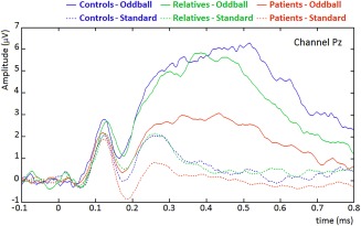
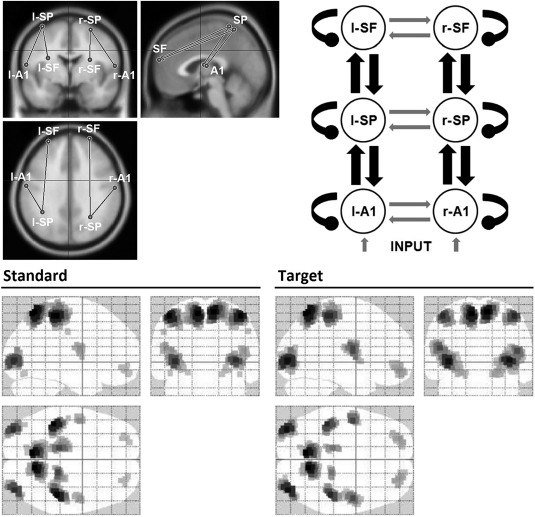
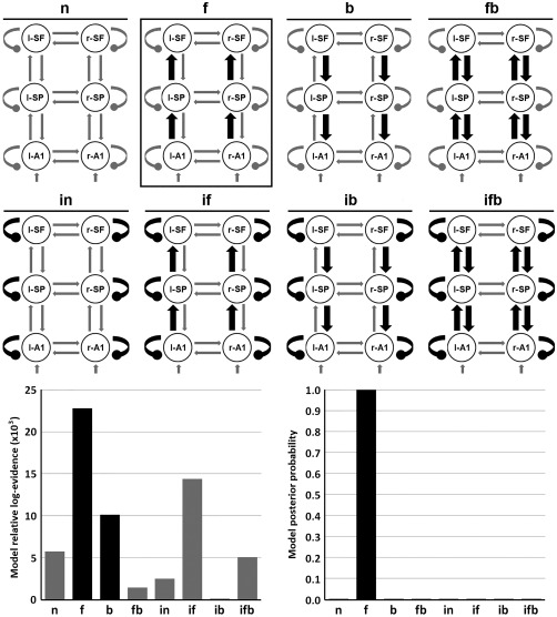
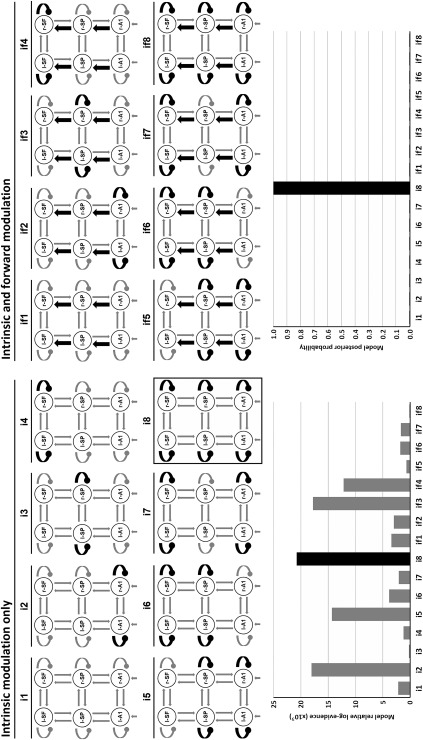
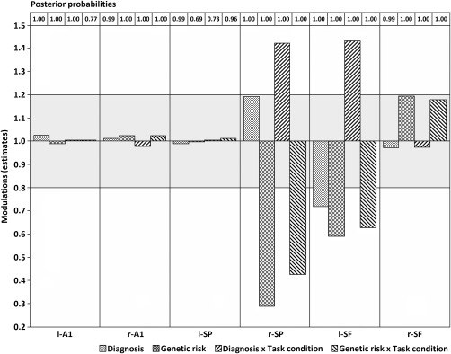
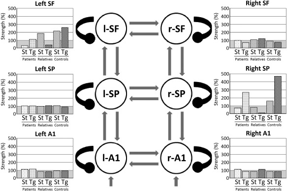
The "dysconnection hypothesis" of psychosis suggests that a disruption of functional integration underlies cognitive deficits and clinical symptoms. Impairments in the P300 potential are well documented in psychosis. Intrinsic (self-)connectivity in a frontoparietal cortical hierarchy during a P300 experiment was investigated. Dynamic Causal Modeling was used to estimate how evoked activity results from the dynamics of coupled neural populations and how neural coupling changes with the experimental factors. Twenty-four patients with psychotic disorder, twenty-four unaffected relatives, and twenty-five controls underwent EEG recordings during an auditory oddball paradigm. Sixteen frontoparietal network models (including primary auditory, superior parietal, and superior frontal sources) were analyzed and an optimal model of neural coupling, explaining diagnosis and genetic risk effects, as well as their interactions with task condition were identified. The winning model included changes in connectivity at all three hierarchical levels. Patients showed decreased self-inhibition-that is, increased cortical excitability-in left superior frontal gyrus across task conditions, compared with unaffected participants. Relatives had similar increases in excitability in left superior frontal and right superior parietal sources, and a reversal of the normal synaptic gain changes in response to targets relative to standard tones. It was confirmed that both subjects with psychotic disorder and their relatives show a context-independent loss of synaptic gain control at the highest hierarchy levels. The relatives also showed abnormal gain modulation responses to task-relevant stimuli. These may be caused by NMDA-receptor and/or GABAergic pathologies that change the excitability of superficial pyramidal cells and may be a potential biological marker for psychosis. Hum Brain Mapp 38:3262-3276, 2017. © 2017 Wiley Periodicals, Inc.
Keywords: DCM; GABA; NMDA; P300; cortical excitability; dynamic causal modeling; effective connectivity; genetic risk; intrinsic connectivity; psychosis; schizophrenia; self-inhibition; synaptic gain; unaffected relatives.
© 2017 Wiley Periodicals, Inc.
Figures
Similar articles
-
Impaired prefrontal synaptic gain in people with psychosis and their relatives during the mismatch negativity.Hum Brain Mapp. 2016 Jan;37(1):351-65. doi: 10.1002/hbm.23035. Epub 2015 Oct 27. Hum Brain Mapp. 2016. PMID: 26503033 Free PMC article.
-
White matter alterations related to P300 abnormalities in individuals at high risk for psychosis: an MRI-EEG study.J Psychiatry Neurosci. 2011 Jul;36(4):239-48. doi: 10.1503/jpn.100083. J Psychiatry Neurosci. 2011. PMID: 21299920 Free PMC article.
-
Association between hippocampal volume and P300 event related potential in psychosis: support for the Kraepelinian divide.Neuroimage. 2012 Jan 16;59(2):997-1003. doi: 10.1016/j.neuroimage.2011.08.067. Epub 2011 Sep 5. Neuroimage. 2012. PMID: 21924362
-
Resting-state EEG source localization and functional connectivity in schizophrenia-like psychosis of epilepsy.PLoS One. 2011;6(11):e27863. doi: 10.1371/journal.pone.0027863. Epub 2011 Nov 18. PLoS One. 2011. PMID: 22125634 Free PMC article.
-
Is auditory processing measured by the N100 an endophenotype for psychosis? A family study and a meta-analysis.Psychol Med. 2024 Jun;54(8):1559-1572. doi: 10.1017/S0033291723003409. Epub 2023 Nov 24. Psychol Med. 2024. PMID: 37997703
Cited by
-
Top-Down Disconnectivity in Schizophrenia During P300 Tasks.Front Comput Neurosci. 2018 May 23;12:33. doi: 10.3389/fncom.2018.00033. eCollection 2018. Front Comput Neurosci. 2018. PMID: 29875646 Free PMC article.
-
Computational Modeling of Electroencephalography and Functional Magnetic Resonance Imaging Paradigms Indicates a Consistent Loss of Pyramidal Cell Synaptic Gain in Schizophrenia.Biol Psychiatry. 2022 Jan 15;91(2):202-215. doi: 10.1016/j.biopsych.2021.07.024. Epub 2021 Aug 10. Biol Psychiatry. 2022. PMID: 34598786 Free PMC article.
-
Different Contexts in the Oddball Paradigm Induce Distinct Brain Networks in Generating the P300.Front Hum Neurosci. 2019 Jan 7;12:520. doi: 10.3389/fnhum.2018.00520. eCollection 2018. Front Hum Neurosci. 2019. PMID: 30666193 Free PMC article.
-
Alterations in Schizophrenia-Associated Genes Can Lead to Increased Power in Delta Oscillations.Cereb Cortex. 2019 Feb 1;29(2):875-891. doi: 10.1093/cercor/bhy291. Cereb Cortex. 2019. PMID: 30475994 Free PMC article.
-
Altered Effective Connectivity within an Oculomotor Control Network in Unaffected Relatives of Individuals with Schizophrenia.Brain Sci. 2021 Sep 17;11(9):1228. doi: 10.3390/brainsci11091228. Brain Sci. 2021. PMID: 34573248 Free PMC article.
References
-
- American Psychiatric Association (1994): Diagnostic and statistical manual of mental disorders : DSM‐IV, 4th ed Washington DC: American Psychiatric Association.
-
- Artigas F (2010): The prefrontal cortex: A target for antipsychotic drugs. Acta Psychiatr Scand 121:11–21. - PubMed
Publication types
MeSH terms
Grants and funding
LinkOut - more resources
Full Text Sources
Other Literature Sources
Medical
Miscellaneous
