Review
. 2017 Feb;65(2):103-108.
doi: 10.4103/ijo.IJO_865_16.

Innovations in glaucoma surgery from Dr. Rajendra Prasad Centre for Ophthalmic Sciences

Affiliations
Review

Innovations in glaucoma surgery from Dr. Rajendra Prasad Centre for Ophthalmic Sciences

Tanuj Dada et al. Indian J Ophthalmol. 2017 Feb.

Abstract

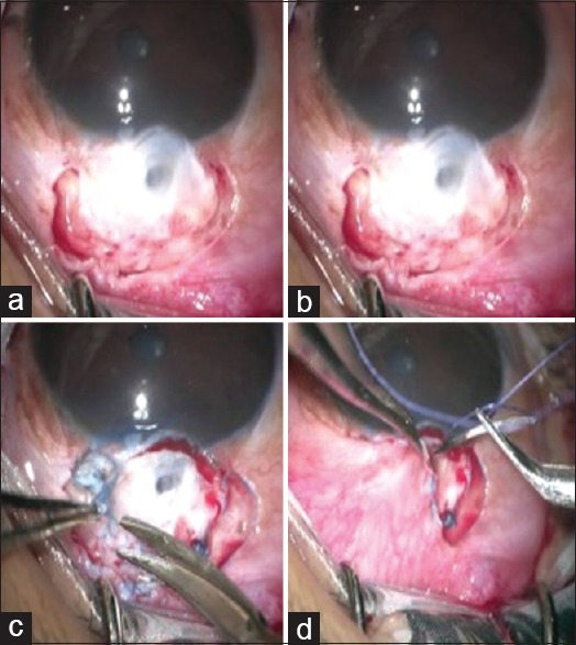
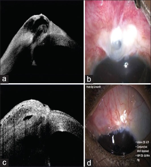
Trabeculectomy surgery is the current standard of care in glaucoma for achieving a low target intraocular pressure if medical therapy is not adequate. Augmentation of trabeculectomy with antimetabolites brought a revolutionary change in the long-term success rates of trabeculectomy, but along with it came a plethora of complications. There still is a big window for therapeutic innovations on this subject. The foremost target for these innovations is to modulate the wound healing response after glaucoma drainage surgery. Achieving the desired balance between long-term success of filtering blebs versus early failure due to scarring of blebs and hypotony due to dysfunctional filtering blebs poses a unique challenge to the ophthalmologists. Alternatives to trabeculectomy such as glaucoma drainage devices and minimally invasive glaucoma surgeries cannot solve the problem of glaucoma blindness in our country, mainly due to their unpredictable results and unfavorable cost-benefit ratio. In this article, we present a summary of our innovations in glaucoma surgery to advance patient care by making it more effective, safer, and economical.


Conflict of interest statement

There are no conflicts of interest.

Figures

Figure 1
Figure 1
(a) Conventional partial thickness scleral flap is raised and two partial thickness vertical cuts are made at the scleral bed. (b) Smaller partial thickness flap is raised from the scleral bed. (c) Smaller flap is cut. (d) A deep scleral pocket is created to allow pooling of aqueous
Figure 2
Figure 2
Postoperative anterior segment optical coherence tomography at 3 months follow-up showing a well-elevated bleb with subconjunctival Ologen (red asterisk) in situ
Figure 3
Figure 3
(a) 40 gauge illuminated microcatheter (DORC international). (b) Schlemm's canal exposed for inserting illuminated microcatheter
Figure 4
Figure 4
(a) Flat bleb with increased vascularity. (b) Corresponding live intraoperative optical coherence tomography imaging-horizontal scan (blue horizontal line) and vertical scan (red vertical line) of the bleb area shows minimal hyporeflective spaces, indicating a fibrosed bleb. (c) Bleb begins to elevate after needling. (d) Intra-operative intraoperative optical coherence tomography imaging showing a relatively raised bleb with multiple hypo-reflective spaces
Figure 5
Figure 5
Bleb sparing epithelial exchange technique of bleb revision. (a) The conjunctiva around the bleb area is separated. (b) The dead epithelium over bleb is stained using trypan blue dye. (c) Stained epithelium is peeled off, leaving underlying bleb intact. (d) The surrounding conjunctiva is advanced over the bleb
Figure 6
Figure 6
(a) Clinical photograph showing thin-walled avascular bleb. (b) Corresponding anterior segment optical coherence tomography showing a thin layer of conjunctiva over the bleb area. (c) Postoperative clinical photograph showing healthy conjunctiva advanced over the bleb. (d) Corresponding anterior segment optical coherence tomography showing well elevated bleb, covered with a healthy conjunctiva

References

    1. Cairns JE. Trabeculectomy. Preliminary report of a new method. Am J Ophthalmol. 1968;66:673–9. - PubMed
    1. Landers J, Martin K, Sarkies N, Bourne R, Watson P. A twenty-year follow-up study of trabeculectomy: Risk factors and outcomes. Ophthalmology. 2012;119:694–702. - PubMed
    1. Lehmann OJ, Bunce C, Matheson MM, Maurino V, Khaw PT, Wormald R, et al. Risk factors for development of post-trabeculectomy endophthalmitis. Br J Ophthalmol. 2000;84:1349–53. - PMC - PubMed
    1. Higashide T, Ohkubo S, Sugimoto Y, Kiuchi Y, Sugiyama K. Persistent hypotony after trabeculectomy: Incidence and associated factors in the Collaborative Bleb-related Infection Incidence and Treatment Study. Jpn J Ophthalmol. 2016;60:309–18. - PubMed
    1. Gedde SJ, Herndon LW, Brandt JD, Budenz DL, Feuer WJ, Schiffman JC. Tube Versus Trabeculectomy Study Group. Postoperative complications in the tube versus trabeculectomy (TVT) study during five years of follow-up. Am J Ophthalmol. 2012;153:804–14.e1. - PMC - PubMed